Source:healthtalk.unchealthcare.org
Diabetic retinopathy is the most common cause of vision loss in people who have diabetes and the leading cause of blindness in working-age people in the United States. It can be prevented, but it is difficult to catch because it usually has no symptoms in the early stages of the disease. Once damage is done, it often cannot be reversed.
As the prevalence of diabetes rises, so does vision loss: In 2010, about 7.7 million Americans had diabetic retinopathy; by 2050, the Centers for Disease Control and Prevention anticipates that number will be 14.6 million.
UNC ophthalmologist Seema Garg, MD, PhD, is working to educate people about the condition and how to prevent the vision loss caused by diabetes.
What Is Diabetic Retinopathy?
Diabetic retinopathy is caused by the effects of diabetes on blood vessels in the retina, the layer of tissue lining the back wall of the eye that sends signals to the brain in response to light and allows us to see.
People with uncontrolled diabetes have excess sugar in the bloodstream, which causes damage to blood vessels throughout the body. In the retina, this excess blood sugar can cause blockages that stop blood flow or produce leaks of fluid and blood. This can result in cloudy or blurred vision that can lead to blindness.
If you have diabetes, you are potentially at risk for developing diabetic retinopathy, which is why a yearly comprehensive eye exam is so important, Dr. Garg says.
“With timely screening and intervention, 90 percent of vision loss from diabetes can be prevented,” Dr. Garg says. “Therefore, guidelines have been established that recommend all patients with diabetes should receive at least an annual retinal exam.”
Diabetic Retinopathy: Symptoms and Diagnosis
Symptoms of diabetic retinopathy may include blurry vision, seeing spots or floaters, having a dark or empty spot in your eyesight, and difficulty seeing well at night. These symptoms usually don’t appear until later stages of the disease, when more damage has been done—and that damage may not be reversible.
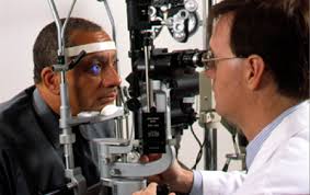
Ophthalmologists can check for diabetic retinopathy several ways. The most common method is with pupil dilation, in which your doctor gives you eye drops to widen the center of the iris of your eye, which makes it easier to see issues in the inner parts of the eye.
If diabetic retinopathy is suspected, your doctor may follow up with retinal photography, which uses a camera to take a picture of the retina, or fluorescein angiography, a procedure in which a fluorescent dye is injected into the bloodstream, making blood vessels easier to photograph.
There are two types of diabetic retinopathy: nonproliferative (NPDR) and proliferative (PDR). NPDR is classified as the early stage of the disease in which there may be vision loss from leaky blood vessels, causing swelling of the retina (diabetic macular edema).
PDR is the more advanced form of diabetic retinopathy, when new, fragile, abnormal blood vessels grow, which may cause significant vision loss from bleeding and retinal detachment. Once diagnosed, treatment can begin to slow or stop the retinal damage caused by diabetes.
Treatment and Prevention of Diabetic Retinopathy
Treatment depends on whether you have NPDR or PDR. In early stages of the disease, your doctor may prescribe regular monitoring, along with a balanced diet to help manage blood sugar levels, exercise and adherence to your medications, including insulin.
For more advanced stages of diabetic retinopathy, laser treatment can be used to stop leakage of blood or fluid in the retina. Medication can also be injected into the eye to help treat diabetic macular edema and prevent new blood vessels from growing and causing more bleeding. In severe cases, your doctor may recommend a surgery called vitrectomy, which removes the vitreous, the gel-like fluid in the eye, and replaces it with another clear substance.
Of course, it is ideal to prevent diabetic retinopathy before it starts. The best way to do this is to prevent the onset of type 2 diabetes by maintaining a healthy weight through exercise and a balanced diet. Some types of diabetes, like type 1 and gestational, cannot be prevented with diet and exercise.
For those diagnosed with diabetes, managing blood sugar levels and receiving a comprehensive yearly eye exam is the best way to prevent diabetic retinopathy or its progression, but for many people that’s not as simple as it sounds.
“Due to a variety of barriers—financial strain, lack of transportation, lack of awareness as to the threat to vision—on average, less than half of all Americans with diabetes receive their yearly retinal screening evaluation,” Dr. Garg says.
Screening for Diabetic Retinopathy
The screening rate for diabetic retinopathy can be as low as 3 percent in rural areas of North Carolina, Dr. Garg says. As a result, rates of vision loss in these communities are high. Through a study, Dr. Garg and fellow UNC physicians are trying to change this. They’re using telemedicine to bring retinal screening to patients during their routine diabetes clinic visits with their primary care doctors. The patients have their retinas evaluated remotely by an ophthalmologist, such as Dr. Garg.
“This is great for patients,” says Tom Miller, MD, a UNC internal medicine physician who is helping conduct the study. “Patients have their exam completed in about 10 minutes while they’re seeing their primary care doctor. We’re capturing people that probably wouldn’t get their eyes evaluated otherwise.”
The study also uses a new type of camera technology that captures a bigger and better image of the retina and does not require pupil dilation. The images are uploaded to a secure website and are remotely interpreted by Dr. Garg. Within 48 hours, a report containing a preliminary diagnosis and management plan is sent to the patient and his or her physician to discuss. If needed, patients with a certain degree of retinopathy are referred to an ophthalmologist for further eye care. Otherwise, the patient is scheduled for a repeat photograph in six months or a year.
