
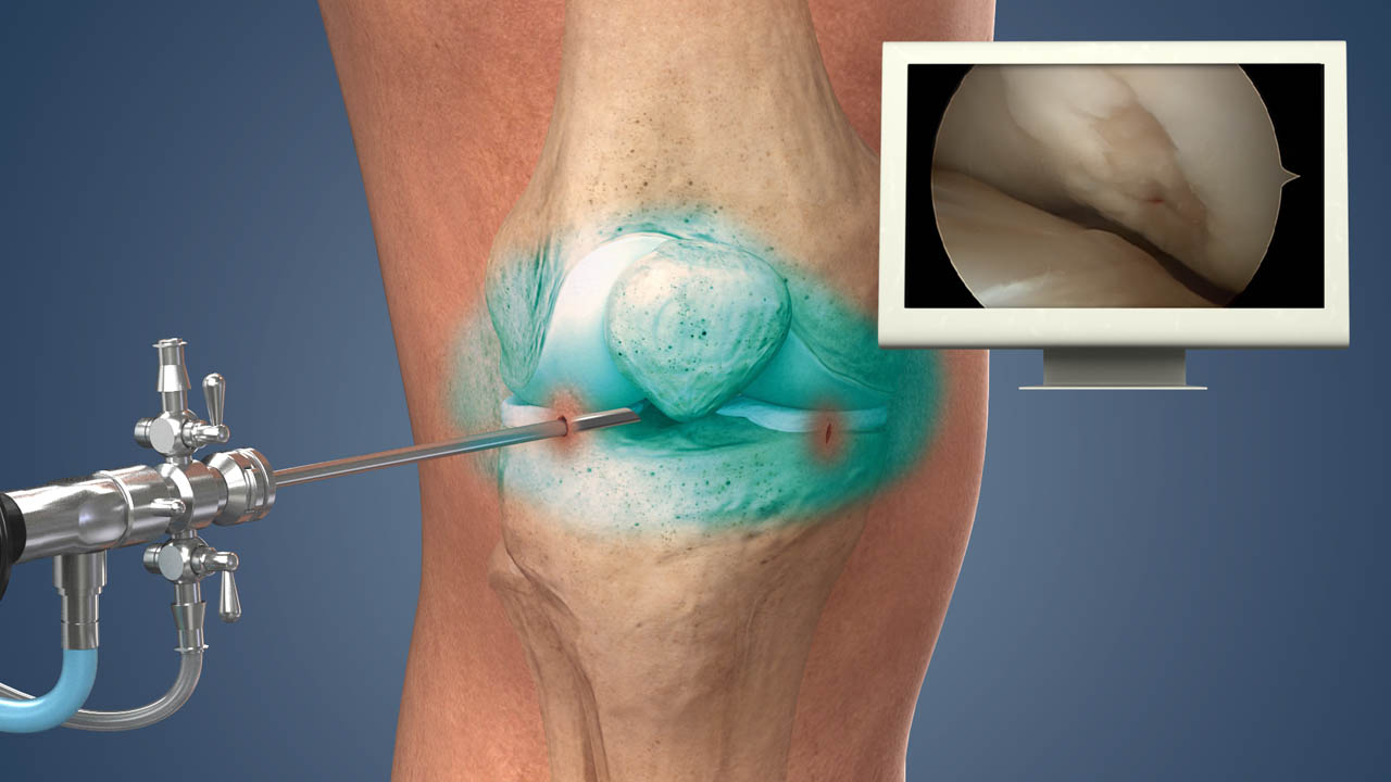
Introduction to Arthroscopic Shaving of Cartilage
Articular cartilage is the smooth, gliding tissue that covers the ends of bones in joints, enabling low-friction, nearly pain-free motion. However, cartilage has limited self-healing capacity because it is avascular, aneural, and lacks a robust supply of reparative cells. Over time, injury, wear, or degeneration can lead to cartilage fraying, fissures, loose flaps, or partial delamination, which may cause pain, swelling, mechanical symptoms (catching, locking), and progression toward osteoarthritis.
Arthroscopic shaving of cartilage-also termed arthroscopic debridement, chondroplasty, abrasion, or cartilage trimming-is a minimally invasive procedure in which damaged or unstable cartilage fragments are removed (or smoothed) via arthroscopic instruments. The idea is not to regenerate full cartilage, but to relieve irritative mechanical symptoms, stabilize cartilage edges, and allow the joint to function more smoothly. In selected patients (especially focal lesions without advanced arthritis), it can provide symptom relief, delay further joint damage, and serve as a bridge to more advanced cartilage repair techniques.
Because the evidence is mixed (especially for osteoarthritic joints), careful patient selection is critical. Arthroscopic shaving is generally considered when symptoms persist despite conservative therapy and the lesion(s) are focal rather than diffuse.
Causes and Risk of Arthroscopic Shaving Of Cartilage
Arthroscopic shaving of cartilage-also known as chondral debridement or chondroplasty-is a minimally invasive procedure performed to smooth rough or frayed articular cartilage inside the knee joint. It is mainly used to reduce pain, remove loose tissue, and improve joint motion when cartilage surfaces have been damaged but not completely worn out.
Here we consider both the underlying etiologies of cartilage damage that might lead to needing shaving, and the risks or factors that influence outcomes of the procedure.
Underlying Causes / Indications (Why cartilage gets damaged)
-
Acute trauma / injury: sports injuries, ligament tears, meniscal tears, chondral impact injuries may create cartilage fissures or flaps
-
Repetitive microtrauma / overuse: overloading, joint malalignment (e.g. varus/valgus), footwear issues
-
Focal osteochondral lesions: e.g. of femoral condyles, talar dome, trochlear cartilage
-
Osteoarthritis / degenerative change: cartilage breakdown, fissuring, surface roughening
-
Meniscal or ligament injuries that change joint mechanics, increasing focal cartilage stress
-
Loose bodies / cartilage flaps that cause mechanical irritation
-
Inflammatory joint disease (in some contexts)
-
Iatrogenic causes (prior surgery altering joint surfaces)
Risk / Prognostic Factors (Factors affecting outcome)
-
Extent of cartilage damage: small, focal lesions do better than diffuse cartilage wear
-
Coexisting joint degeneration (arthritis) - patients with advanced osteoarthritis generally have less benefit
-
Patient age: younger patients tend to respond better
-
Body mass index (BMI) / obesity: more load on joint may reduce benefit
-
Alignment / mechanical axis: malalignment (varus/valgus) or instability can continue to stress cartilage
-
Concomitant pathology: meniscal tears, ligament injuries, maltracking
-
Joint environment (synovitis, inflammation)
-
Duration of symptoms
-
Postoperative compliance and rehabilitation
Because shaving only removes unstable cartilage flaps and smooths rough surfaces (without replacing cartilage), success depends heavily on the underlying cartilage environment and joint mechanics.
Symptoms and Signs of Cartilage Lesions (Where Shaving Might Be Indicated)
Cartilage lesions-localized areas of cartilage damage within the knee-present with mechanical and inflammatory symptoms that interfere with smooth joint movement. Recognizing these signs early is essential, as such lesions often lead to procedures like arthroscopic shaving (chondroplasty) when conservative treatment fails.
Patients who may be candidates for arthroscopic cartilage shaving often present with:
-
Joint pain (aching, sharp, intermittent) - often with weight-bearing or activity
-
Mechanical symptoms such as catching, locking, clicking
-
Swelling or joint effusion
-
Tenderness localized to the affected joint area
-
Occasional giving way or instability sensation (especially if associated injuries)
-
Pain on motion, crepitus or rough sensation internally during movement
-
Reduced range of motion (particularly at extremes)
-
Functional limitation - difficulty in stair-climbing, squatting, twisting, pivoting
On physical exam:
-
Local joint line tenderness
-
Pain or crepitus on movement
-
Swelling / effusion
-
Assessment of alignment, ligament stability, meniscus, gait
-
Range of motion testing - both active and passive
These signs are nonspecific, so imaging and arthroscopic evaluation are generally needed to localize and quantify the cartilage damage.
Diagnosis of Arthroscopic Shaving Of Cartilage
The diagnosis for arthroscopic shaving of cartilage involves a combination of clinical evaluation, imaging findings, and direct visualization through arthroscopy, which serves both as a diagnostic and therapeutic tool. It is recommended when other diagnostic methods confirm focal cartilage injury but conservative treatments fail to relieve symptoms.
Imaging Modalities
-
Plain radiographs (X-ray): AP, lateral, and special views to assess joint space narrowing, osteophytes, subchondral changes
-
Weight-bearing radiographs to assess load-bearing joint surfaces
-
MRI (Magnetic Resonance Imaging): the preferred noninvasive tool for cartilage imaging; can detect cartilage defects, subchondral bone changes, associated meniscal or ligament pathology
-
MRI cartilage mapping / advanced sequences (e.g., T2 mapping, dGEMRIC) may help assess cartilage quality
-
CT / CT arthrography in selected cases (especially to assess subchondral bone, depth, defect geometry)
-
Ultrasound (limited in cartilage)
-
Bone scan / SPECT-CT (rarely) if needed to evaluate bone metabolism
Modern methods, including deep learning applied to MRI, are being explored to improve sensitivity for detecting cartilage defects.
Diagnostic Arthroscopy
-
Arthroscopy remains the gold standard for visualizing cartilage lesions directly, assessing their stability, size, depth, and adjacent joint surfaces
-
During arthroscopy, surgeons can probe cartilage flaps, assess viability, and decide on shaving / debridement vs repair
Clinical & Preoperative Evaluation
-
Thorough history (duration, aggravating/relieving factors, mechanical symptoms)
-
Physical examination (alignment, ligament / meniscal assessment, adjacent structures)
-
Assessment of comorbidities (e.g. obesity, metabolic disease)
-
Assessment of joint environment (synovitis, swelling)
-
Counseling patient about expectations (shaving is palliative, not restorative)
The decision to proceed with arthroscopic shaving should take into account the lesion's characteristics, patient factors, and alternative repair options.
Treatment Options Role of Arthroscopic Shaving
Arthroscopic shaving (chondroplasty or debridement) is a minimally invasive treatment option for cartilage lesions designed to relieve pain, smooth irregular surfaces, and remove loose tissue within the joint. It forms part of a broader set of cartilage restoration and preservation techniques that range from palliative to restorative, depending on lesion severity and patient condition.
Conservative / Non-surgical Management (Before Considering Shaving)
Before surgical intervention, many patients try:
-
Activity modification / avoiding aggravating activities
-
Pain management: NSAIDs, analgesics
-
Physical therapy: strengthening, proprioception, muscle balancing
-
Bracing, orthoses to offload
-
Injections: corticosteroids, hyaluronic acid, platelet-rich plasma (PRP) in some settings
-
Weight loss, lifestyle modification
-
Nutritional support, cartilage-protecting supplements (though evidence is limited)
If conservative therapy fails to relieve symptoms sufficiently, surgical options may be considered.
Arthroscopic Shaving / Debridement (the focus intervention)
What it does:
-
Remove loose cartilage flaps, fibrillated tissue, unstable edges (shaving or smoothing)
-
Achieve stable, smooth cartilage margins (chondroplasty)
-
Optionally perform adjunctive smoothing or abrasion of subchondral bone (carefully)
-
May relieve pain, reduce irritation from debris, improve mechanical environment
When it is indicated (favorable scenarios):
-
Focal cartilage lesions with unstable flaps
-
Minimal or no advanced osteoarthritis
-
Patients with mechanical symptoms (catching, locking)
-
Good joint alignment and stable biomechanics
-
When cartilage repair options are not feasible or as a temporizing measure
Limitations:
-
Does not regenerate cartilage - it is a palliative / symptomatic procedure
-
Less effective in diffuse or high-grade cartilage degeneration
-
Benefits may diminish over time if underlying degenerative process continues
Evidence & Outcomes:
-
A systematic review of arthroscopic cartilage debridement in the knee (focal lesions) reported good to excellent short- and medium-term functional results in selected patients.
-
However, debridement in the setting of osteoarthritis (diffuse disease) has shown conflicting (often disappointing) long-term benefits; some trials suggest limited efficacy versus placebo or nonoperative therapy.
-
In ankle/talar lesions (e.g. osteochondral lesions of talus), arthroscopic debridement (alone or with adjuncts) has been used with reasonable short-to-mid-term success, though long-term data are less robust.
Because of these limitations, surgeons often combine shaving with cartilage repair techniques (microfracture, drilling, osteochondral grafting, cell-based methods) when feasible.
Adjunct or Alternative Cartilage Repair Techniques
When cartilage repair or restoration is the goal (rather than mere symptomatic relief), surgeons may employ:
-
Microfracture / drilling / bone marrow stimulation - to encourage fibrocartilage repair
-
Osteochondral autograft / allograft transplantation (OATS, mosaicplasty)
-
Autologous chondrocyte implantation (ACI) / matrix-assisted ACI (MACI)
-
Minced cartilage implantation / cartilage scaffolds / matrices (single-stage arthroscopic techniques)
-
Biologic adjuncts (e.g. PRP, growth factors, stem cell therapies)
-
Scaffolds / hydrogels / biomaterials
In practice, shaving may serve as the first step (to clear unstable tissue) before applying these advanced repair methods in a hybrid approach.
Prevention and Management (Before & After Procedure)
Prevention and management surrounding arthroscopic cartilage shaving focus on protecting knee health before surgery and ensuring optimal recovery and joint preservation afterward. The goals are to reduce pain, restore smooth joint motion, and prevent further cartilage degeneration.
Prevention / Before Damage
-
Avoid repetitive overload, especially in malaligned joints (correct alignment when possible)
-
Use proper footwear, joint-protective devices
-
Maintain healthy weight / reduce obesity
-
Strengthening and conditioning of muscles around joints
-
Treat joint injuries early (meniscus, ligament) to prevent secondary cartilage damage
-
Maintain joint health (nutrition, controlling metabolic diseases)
Preoperative Optimization
-
Ensure good mechanical alignment / stability
-
Address concomitant lesions (ligament, meniscus)
-
Manage comorbidities (obesity, diabetes, etc.)
-
Patient education: realistic expectations, rehab compliance
Postoperative / Rehabilitation & Management
-
Early mobilization (within limits) and controlled range-of-motion exercises
-
Gradual return to weight-bearing as tolerated (per surgeon's protocol)
-
Physical therapy focusing on strength, proprioception, joint motion
-
Activity modification (avoid high-impact for a period)
-
Use of orthoses or braces if needed
-
Periodic follow-up (clinical + imaging)
-
Monitoring for recurrence or progression, and considering further repair if needed
Patient compliance, gradual progression, and controlling mechanical stresses are key to long-term benefit.
Complications of Arthroscopic Shaving (Debridement / Chondroplasty)
Arthroscopic shaving (chondroplasty/debridement) is generally considered safe, but complications can occur, with an overall rate of about 3.5–3.6% based on large studies. Most complications are surgical, with infection being the most common, and the risk profile is similar or slightly higher compared to meniscectomy.
-
Persistent or recurrent pain / symptoms (i.e. failure to relieve symptoms)
-
Worsening of cartilage damage / progression of degenerative joint disease
-
Iatrogenic cartilage or cartilage-edge injury from instrumentation
-
Subchondral bone damage / bleeding / thermal injury (especially if using radiofrequency / thermal devices)
-
Joint swelling, effusion, synovitis
-
Infection (rare in arthroscopy)
-
Hemarthrosis (bleeding into joint)
-
Neurovascular injury (rare)
-
Thromboembolic complications (rare)
-
Stiffness / limited motion
-
Need for revision procedures or cartilage repair/grafting
Surgeons must weigh the risk-benefit, especially in borderline cases, and counsel patients appropriately.
Living With the Condition / Post-operative Outlook & Patient Guidance
After cartilage shaving, patients must adapt and optimize joint health. Realistic expectations are crucial: shaving is unlikely to restore perfect cartilage but aims to relieve symptoms and delay progression.
Typical Outcomes & What Patients Can Expect
-
Many patients experience improvement in pain and function in the short to medium term, particularly for focal lesions
-
The benefit may diminish over time if degeneration continues
-
In well-selected cases, shaving can serve as a bridge to more definitive cartilage repair
-
Some patients may still require further interventions later
Tips & Lifestyle Guidance
-
Use low-impact activities (swimming, cycling) rather than high-impact ones (running) initially
-
Wear good supportive footwear and insoles / orthoses to distribute load
-
Maintain healthy weight to reduce stress
-
Continue strengthening and flexibility exercises to support joint mechanics
-
Monitor the joint (pain, swelling, new mechanical symptoms) and seek evaluation if symptoms recur
-
Adhere strictly to rehabilitation protocols
-
Stay under periodic orthopedic follow-up
Long-Term Monitoring & Possible Next Steps
-
Use follow-up imaging (MRI, arthroscopy if needed) to gauge cartilage environment
-
If symptoms progress or cartilage repair fails, consider cartilage restoration techniques (e.g. microfracture, grafting, cell therapy)
-
In advanced joint degeneration, joint replacement or fusion may eventually be considered
Top 10 Frequently Asked Questions about Arthroscopic Shaving of Cartilage
1. What is Arthroscopic Shaving of Cartilage?
Arthroscopic shaving of cartilage, also known as chondroplasty, is a minimally invasive surgical procedure performed to smooth damaged cartilage surfaces in joints, most commonly the knee. It is used to relieve pain, improve joint function, and reduce mechanical symptoms such as catching or locking caused by cartilage defects.
2. Why is Arthroscopic Shaving of Cartilage Performed?
The procedure is recommended for patients with:
-
Cartilage tears or fraying due to injury or degenerative conditions.
-
Osteoarthritis causing rough cartilage surfaces.
-
Mechanical symptoms like joint locking, catching, or clicking.
-
Pain and limited range of motion not relieved by conservative treatments like medications, physiotherapy, or injections.
The goal is to restore smooth joint surfaces, reduce pain, and improve mobility.
3. How is Arthroscopic Shaving of Cartilage Performed?
The procedure involves:
-
Anesthesia: General or regional anesthesia.
-
Small Incisions: Typically 2-3 tiny incisions around the joint.
-
Arthroscope Insertion: A small camera is inserted to visualize the joint.
-
Shaving or Smoothing: Damaged cartilage is trimmed and smoothed using specialized instruments.
-
Closure: Incisions are closed with sutures or steri-strips, and a sterile dressing is applied.
The procedure usually takes 30-60 minutes and is often performed as an outpatient surgery.
4. What Are the Benefits of Arthroscopic Shaving of Cartilage?
Key benefits include:
-
Pain relief by removing rough cartilage surfaces.
-
Improved joint function and range of motion.
-
Minimally invasive with smaller scars and quicker recovery.
-
Reduced mechanical symptoms such as catching or locking.
-
Shorter downtime compared to open joint surgery.
5. Who is an Ideal Candidate for the Procedure?
Ideal candidates include:
-
Adults with localized cartilage damage or fraying.
-
Patients experiencing pain, swelling, and mechanical symptoms.
-
Individuals who have failed conservative treatment methods.
Patients with advanced osteoarthritis or widespread cartilage loss may require alternative treatments, such as joint replacement.
6. What is the Recovery Process After Surgery?
Recovery typically involves:
-
Immediate post-op: Ice, elevation, and limited activity to reduce swelling.
-
Weight Bearing: Depending on the joint and extent of cartilage damage, partial or full weight bearing may be allowed immediately or after a short period.
-
Physical Therapy: Exercises to restore strength, flexibility, and joint function are initiated within days to weeks.
-
Return to Activities: Most patients resume daily activities within 1-4 weeks, depending on the joint and extent of surgery.
7. Are There Any Risks or Complications?
Although generally safe, risks may include:
-
Infection at the incision site or joint (rare)
-
Blood clots (deep vein thrombosis)
-
Persistent pain or swelling
-
Stiffness or reduced range of motion
-
Rarely, injury to surrounding cartilage, ligaments, or nerves
Following post-operative care and attending physical therapy reduces the likelihood of complications.
8. How Long Do the Benefits Last?
The benefits depend on the extent of cartilage damage and underlying joint health:
-
Many patients experience pain relief and improved function for months to years.
-
In degenerative conditions like osteoarthritis, cartilage deterioration may continue, and additional treatments may be required in the future.
-
Maintaining a healthy weight, regular exercise, and joint protection strategies help prolong results.
9. Can Arthroscopic Shaving of Cartilage Be Combined with Other Procedures?
Yes. The procedure is often combined with:
-
Meniscus repair or partial meniscectomy
-
Synovial debridement to remove inflamed tissue
-
Microfracture or cartilage regeneration techniques in certain cases
-
Ligament reconstruction if associated injuries are present
Combining procedures can address multiple joint issues simultaneously and optimize outcomes.
10. How Can I Prepare for Arthroscopic Shaving of Cartilage?
Preparation typically includes:
-
Medical evaluation: Blood tests, imaging (X-ray or MRI) to assess cartilage damage.
-
Medication review: Especially blood thinners or anti-inflammatory drugs.
-
Pre-operative exercises: Strengthening surrounding muscles may improve recovery.
-
Planning post-surgery care: Assistance at home may be needed for a few days.
Following your surgeon's specific pre-operative instructions ensures a safe procedure and smooth recovery.
