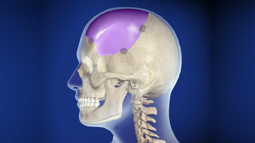
Introduction to Cranioplasty
Cranioplasty is a surgical procedure performed to repair or reconstruct a defect or deformity in the skull - typically the result of prior trauma, surgery (such as a decompressive craniectomy), congenital skull abnormality, or a disease process. The term comes from "cranio-” (skull) and "-plasty” (shaping/repair). In essence, the aim of cranioplasty is threefold: (1) to protect the underlying brain by restoring the bony skull vault, (2) to restore normal skull contour and cosmetic appearance, and (3) to restore more normal intracranial dynamics (pressure, cerebrospinal fluid flow, brain blood flow) that may have been disrupted by the skull defect.
The procedure can employ the patient's own bone flap (autologous bone), or synthetic implants (such as titanium mesh, hydroxyapatite, polymethylmethacrylate, polyetheretherketone) depending on the situation, defect size, timing and condition of the bone.
Cranioplasty is often done a weeks to months after the initial skull-removing surgery (craniectomy) or trauma, once the acute phase of brain swelling or intracranial disease has stabilized. It is considered not just cosmetic, but functional and protective.
In the coming sections, we'll explore how and why cranioplasty is performed, the risks and benefits, how it's diagnosed, treatment approaches, prevention/management, complications, and how patients live with it.
Causes and Risk Factors of Cranioplasty
In this section we examine what leads to the need for cranioplasty, and the risk factors associated with needing this procedure or its complications.
Causes (i.e., reasons why a skull defect exists needing repair)
-
A major cause is a prior decompressive craniectomy (surgical removal of skull bone) done in the setting of traumatic brain injury, stroke, intracranial hemorrhage, or raising intracranial pressure. The resulting skull defect later requires cranioplasty.
-
Trauma or head injury: skull fractures or bone loss from accident, penetrating injury, war injury.
-
Previous neurosurgical procedures: removal of bone flap during tumour resection, infection-related bone removal, bone necrosis.
-
Congenital skull defects or developmental abnormalities of the skull.
-
Bone flap resorption or infection after the first surgery: sometimes the removed bone cannot be replaced and a synthetic implant becomes necessary.
Risk Factors (for needing cranioplasty and for complications)
-
Large size of skull defect: the bigger the bony portion missing, the greater the complexity of repair.
-
Duration of skull defect: longer time between the initial bone removal and reconstruction may increase risks (e.g., bone flap resorption, scalp thinning).
-
Presence of infection or poor wound healing at the time of reconstruction.
-
Poor general health: comorbidities such as diabetes, malnutrition, smoking, steroid use may impair healing.
-
Previous bone flap stored poorly or devitalised: autologous bone flaps may resorb or get infected if storage/handling is suboptimal.
-
Material used for reconstruction: some synthetic materials carry higher risk of infection or rejection than others.
-
Age: children may have particular issues (skull growth, bone resorption) and elderly may have frailer tissues.
Recognising these underlying causes and risk factors allows surgeons and care teams to plan timing, materials and patient optimisation ahead of cranioplasty.
Symptoms and Signs of Cranioplasty
While technically the procedure (cranioplasty) itself is the intervention, in this section we discuss what symptoms or signs might lead to needing a cranioplasty (i.e., problems caused by skull defect) and what patients might experience before and after.
Symptoms/Signs due to skull defect
-
Visible skull indentation or sunken area ("scalp flap sinking”) - sometimes called the "syndrome of the trephined” or "sunken-flap syndrome”. This may lead to headaches, dizziness, intolerance to vibration, fatigue, mood changes, cognitive slowing.
-
Neurological symptoms: because absence of the skull bone may alter brain hemodynamics and cerebrospinal fluid flow, patients may report new or worsening neurological deficits, reduced cognitive function, fatigue or memory issues.
-
Cosmetic/psychosocial: the skull defect may be visible, leading to cosmetic deformity, self-consciousness, social withdrawal.
Signs (clinical findings)
-
On examination: depressed scalp over defect area, palpable edges of bone, sometimes movement of scalp when patient leans forward, visible skull contour irregularity.
-
In imaging: brain CT/MRI may reveal skull defect, change in brain shape, midline shift or localised edema or sinking flap effect.
-
After cranioplasty: one expects the restoration of normal skull contour, improvement of symptoms, fewer headaches, better neurological stability and improved protection of the brain.
By understanding these signs/symptoms, clinicians determine the timing for cranioplasty and address patient-reported issues (both functional and aesthetic).
Diagnosis of Cranioplasty
This section covers how the need for cranioplasty is assessed (diagnosis), how planning is done, and what investigations/surgical planning are involved.
Assessment of skull defect
-
Clinical examination: assessment of skull defect size and location, scalp condition, previous surgery details (bone flap used or not, storage, infection history), neurological status.
-
Review of prior operative notes: to know whether bone flap was preserved, what materials were used, what prior complications occurred.
Imaging studies
-
CT scan of the head (non-contrast) with bone window: to visualise the skull defect, size, margins, condition of bone edges.
-
3D reconstruction/CT bone algorithm: important for custom implant planning.
-
MRI may also be used to evaluate associated brain injury, swelling, CSF flow, and potential contraindications (such as residual brain swelling or hydrocephalus) which may delay cranioplasty.
Preoperative planning
-
Determine the appropriate material for reconstruction (autologous bone vs synthetic implant) based on defect size, condition of bone flap, patient factors (age, comorbidities).
-
Check for infections: ensure the scalp incision and bone flap or storage site are clean; rule out osteomyelitis of bone flap, sinus communication.
-
Evaluate timing: ensure that brain swelling has subsided, infection free, patient is medically stable. Optimal timing remains debated, but many centres wait until the patient is stable and free of infection.
-
Multidisciplinary input: neurosurgery, plastic/reconstructive surgery (especially for complex skull defects), and sometimes maxillofacial surgery may be involved.
In short, diagnosis in this context is more about decision-making and surgical planning rather than diagnosing a disease; yet it is critical to achieve good outcomes.
Treatment Options of Cranioplasty
Here we discuss how cranioplasty is performed, the materials used, timing considerations, operative steps, and postoperative care.
Materials & methods of reconstruction
-
Autologous bone flap: if the previously removed bone flap was preserved (in freezer or in subcutaneous pouch) and remains viable, this may be reused. Advantages: biocompatible, low immunologic risk, lower cost. Challenges: bone resorption, infection risk, bone may be damaged.
-
Synthetic implants: many materials are available. Examples include titanium plates/meshes, polymethylmethacrylate (PMMA), hydroxyapatite, polyetheretherketone (PEEK). These may be prefabricated to fit the skull defect precisely based on preoperative 3D imaging. Materials differ in strength, infection resistance, cost, imaging compatibility.
-
Material choice depends on defect size, patient age, cost, hospital resources and surgeon preference.
Timing of surgery
-
The timing of cranioplasty after the initial defect (e.g., after craniectomy) is somewhat controversial. Some studies suggest earlier reconstruction (< 3 months) may improve neurological recovery and reduce 'sinking flap' complications; others suggest waiting to reduce infection risk.
-
Surgeons aim to operate once brain swelling has resolved, the patient is stable, there is no infection, hydrocephalus is managed.
Operative steps
-
The patient is placed under general anaesthesia. The scalp incision is made (often along previous scar) and scalp and muscle are reflected to expose the defect.
-
The defect edges are cleaned, the implant (or bone flap) is positioned and secured (with plates/screws if synthetic, or fixation devices if bone) to the surrounding skull.
-
Ensuring good scalp coverage, hemostasis, and closure. Sometimes drains or suction are used to prevent fluid collection.
Postoperative care
-
Monitoring in neurosurgical or intensive care setting for early complications (bleeding, infection, fluid collection, neurological status).
-
Imaging (postoperative CT) to check implant position, exclude hematoma.
-
Guidance on physical activity, head protection (if large defect remains), wound care.
-
Rehabilitation as needed (especially if the patient had prior neurological injury).
Goals of cranioplasty
-
Restore skull protection for brain and reduce risk of external trauma.
-
Improve symptoms associated with skull defect (headaches, sinking scalp, neurological symptoms).
-
Improve aesthetic appearance and patient quality of life.
-
Improve cerebrovascular dynamics and intracranial pressure equilibrium. Studies report improvements in cerebral blood flow, CSF circulation and neurological function after cranioplasty.
Prevention and Management of Cranioplasty
This section covers how to reduce the need for cranioplasty complications, how to manage patients before and after the procedure, and preventative strategies.
Preventive strategies before cranioplasty
-
Ensure any infection (systemic or local) is resolved before reconstruction.
-
Optimize general health: control diabetes, hypertension, nutritional status; stop smoking; encourage good skin/scalp condition.
-
Preserve bone flaps carefully if autologous bone is used: proper storage (deep freezing or subcutaneous pouch) and accurate documentation.
-
Plan timing appropriately: delay if brain swelling or hydrocephalus persists, or scalp condition is poor.
Management after cranioplasty
-
Wound care: keep the incision clean, monitor for signs of infection (redness, discharge, fever).
-
Monitor for neurological changes: headaches, seizures, scalp bulge/indentation, fluid collection.
-
Head protection: in some cases where bone implant is fresh or soft tissues thin, using helmet or limiting head trauma may be advised until full healing.
-
Rehabilitation: if the patient had prior brain injury, integrate cranioplasty into overall neurorehabilitation plan (physio, occupational, speech therapy).
-
Follow-up imaging: periodic scans to ensure implant stability, absence of fluid collections, bone resorption (for autologous bone), or implant migration.
-
Patient education: advise on symptoms to watch (infection, wound problems, new neurological deficits), lifestyle modifications (avoid head injury, maintain healthy vascular status).
Through careful preventive management and post-operative follow-up, many complications can be minimised and outcomes optimised.
Complications of Cranioplasty
While cranioplasty is often beneficial, it is not without risks. It is important for patients and caregivers to understand potential complications and how they are managed.
Common complications
-
Infection: The most frequent complication. It may affect the scalp, bone flap or synthetic implant; may require antibiotics and sometimes removal of implant.
-
Hematoma / fluid collection: Subgaleal or subdural collections may form under the implant/scalp, requiring drainage.
-
Bone flap resorption (in autologous bone) - particularly in younger patients or in poorly vascularised bone.
-
Implant failure or displacement/migration: Synthetic implants may shift, especially if fixation is inadequate.
-
Seizures: Potential risk due to prior brain injury, skull defect, or surgery.
-
Neurological deterioration: Rare but possible if underlying brain injury worsens or if there is worsening of CSF/ICP dynamics.
Less common but serious complications
-
Stroke or cerebral infarction (rare)
-
Wound necrosis or skin flap breakdown leading to implant exposure
-
Implant rejection or allergic reaction (rare)
-
Hydrocephalus (fluid accumulation) may require shunt placement or other management; presence of shunt may complicate cranioplasty.
Factors influencing complication risk
-
Timing of cranioplasty (earlier vs delayed) may influence infection risk and neurological recovery - evidence remains mixed.
-
Material used: some studies show lower infection/resorption rates with titanium or custom-made implants compared with some other materials.
-
Patient factors: age, comorbidities, scalp condition, prior radiation, bone flap condition, presence of hydrocephalus or shunt.
Awareness of these risks enables informed consent, vigilant intra- and post-operative care, and early recognition and management of complications.
Living with the Condition of Cranioplasty
This section focuses on the patient journey after cranioplasty: recovery, rehabilitation, long-term outcomes, lifestyle, and expectations.
Recovery and rehabilitation
-
After surgery, most patients stay in hospital for monitoring (usually 2-5 days, longer if complications or prior neurological deficits).
-
Headache, fatigue and mild discomfort are common; pain is managed and activity gradually increased. Walking, physiotherapy, and occupational therapy begin early if needed.
-
Full recovery may take several weeks to months, depending on the patient's prior condition, extent of skull defect, and neurological injury.
Quality of life and functional outcomes
-
Many patients experience significant improvements in symptoms that were due to skull defect - for example less headache, improved cognitive clarity, improved mood, better physical comfort from not having a depressed scalp.
-
Cosmetic restoration can positively affect self-esteem, social interaction and psychological wellbeing.
-
From a functional standpoint, restoration of skull integrity helps protect the brain from external trauma and may improve intracranial physiology (better blood flow and CSF circulation).
Lifestyle considerations
-
Patients are encouraged to maintain general health: healthy diet, stay hydrated, stop smoking, control blood pressure and diabetes, protect head from injury (especially in early recovery).
-
Activities: heavy contact sports or activities with head trauma risk may need modification until surgeon clearance.
-
Headgear/protection: in cases of large defects or thin scalp coverage, helmet use may be advised until full healing.
Long-term follow-up
-
Routine neurosurgery follow-ups: checking for wound healing, implant stability, imaging if needed.
-
Monitor for late complications: scalp thinning, implant exposure, bone resorption or implant failure.
-
Psychological support: if there has been prior brain injury, rehabilitation often includes neuropsychological support for cognitive changes, mood disorders or post-traumatic stress.
Realistic expectations and patient education
-
It's important that patients understand that cranioplasty is not just cosmetic - it has protective and functional benefits.
-
Recovery is individual: patients with prior brain injury may have residual deficits independent of skull repair.
-
Education about signs of complications (e.g., increased headaches, swelling, fever, fluid leak) empowers patients to alert doctors early.
-
Engagement in rehabilitation, healthy lifestyle and protective behaviours optimise long-term outcomes.
Top 10 Frequently Asked Questions about Cranioplasty Surgery
1. What Is Cranioplasty?
Cranioplasty is a surgical procedure to repair or reconstruct a
defect in the skull.
It is typically performed after a craniectomy (when a portion of the
skull is removed to relieve brain pressure) or due to trauma, congenital defects, or
bone loss from infection or tumor removal.
The procedure restores:
-
Protection to the brain
-
Skull contour and cosmetic appearance
-
Normal intracranial pressure dynamics
Cranioplasty may involve using autologous bone (the patient's own bone) or synthetic materials such as titanium, PMMA, or PEEK.
2. Why Is Cranioplasty Performed?
Cranioplasty is recommended for several reasons:
-
To replace bone removed during previous craniectomy
-
To protect the brain and prevent injury
-
To correct skull deformities or asymmetry
-
To restore neurological function, as skull defects can sometimes impair cognition or motor function
-
For cosmetic improvement after trauma or surgery
By reconstructing the skull, cranioplasty enhances both safety and appearance.
3. Who Is a Candidate for Cranioplasty?
Candidates typically include patients who:
-
Have undergone previous craniectomy and now require bone replacement
-
Have skull defects from trauma, tumors, or congenital conditions
-
Are healthy enough to undergo surgery under general anesthesia
-
Do not have active infections at the surgical site
-
Desire cosmetic restoration of skull shape
Your neurosurgeon will evaluate your bone defect, overall health, and timing to determine suitability.
4. How Is Cranioplasty Surgery Performed?
Cranioplasty is performed under general anesthesia.
Procedure Steps:
-
The surgeon reopens the previous incision to expose the skull defect.
-
Any scar tissue or residual debris is removed.
-
The bone flap or synthetic implant is carefully shaped to fit the defect.
-
The implant is secured using plates, screws, or adhesives.
-
The scalp is sutured over the implant, and a dressing is applied.
The procedure typically takes 1–3 hours, depending on the size and complexity of the defect.
5. What Types of Materials Are Used for Cranioplasty?
Cranioplasty can use autologous or synthetic materials:
-
Autologous Bone: The patient's own bone, often preserved from a previous craniectomy.
-
Titanium: Strong, durable metal used for large defects.
-
PMMA (Polymethylmethacrylate): Lightweight, moldable acrylic.
-
PEEK (Polyetheretherketone): Biocompatible, lightweight polymer for custom implants.
The choice depends on defect size, location, risk of infection, and patient preference.
6. What Is the Recovery Process After Cranioplasty?
Recovery varies based on patient health and defect size:
-
Hospital stay: Usually 3–5 days for monitoring, longer if complications arise.
-
Pain and swelling: Mild to moderate, managed with medications.
-
Activity restrictions: Avoid heavy lifting, strenuous exercise, or contact sports for 4–6 weeks.
-
Follow-up: Regular visits to monitor wound healing and implant integration.
Most patients can resume normal activities within a few weeks, but full recovery may take 2–3 months.
7. Are There Any Risks or Complications?
While cranioplasty is generally safe, potential risks include:
-
Infection at the surgical site
-
Bleeding or hematoma formation
-
Implant rejection or displacement
-
Seizures or neurological changes (rare)
-
Scarring
-
Persistent swelling or pain
Most complications are preventable with proper surgical technique, sterile handling, and close follow-up care.
8. How Long Do Cranioplasty Implants Last?
Cranioplasty implants are long-lasting:
-
Autologous bone may integrate with surrounding tissue and last a lifetime but can resorb in some cases.
-
Titanium and synthetic implants are durable and designed to remain permanently in place.
Patients usually require long-term follow-up to ensure implant stability and monitor for any late complications.
9. Will Cranioplasty Affect Brain Function?
Cranioplasty itself is not harmful to brain function. In fact, restoring skull integrity can sometimes improve neurological function:
-
Patients report improvements in cognition, coordination, and motor skills after skull reconstruction.
-
Brain protection is restored, reducing the risk of injury or trauma.
The outcome depends on the underlying brain condition and timing of surgery.
10. What Is the Long-Term Outlook After Cranioplasty?
The long-term prognosis is excellent when performed under proper surgical conditions:
-
Most patients regain normal skull contour and protection.
-
Neurological function and cognitive performance often improve over time.
-
Cosmetic results are usually satisfactory, enhancing confidence.
-
Lifelong follow-up is recommended for monitoring implant integrity and scalp health.
With modern materials and techniques, cranioplasty is highly effective and durable, offering both functional and aesthetic benefits.


