
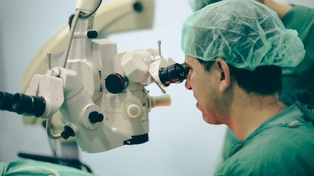
Introduction to Bleb Revision
A "bleb" in ophthalmology refers to the fluid-filled elevation of the conjunctiva created intentionally in a procedure called Trabeculectomy, a surgery for glaucoma that diverts aqueous humour out of the eye to lower intra-ocular pressure (IOP). Over time, however, this filtering bleb may fail, leak, become dysfunctional, or cause discomfort. Bleb revision is the general term for surgical or procedural interventions aimed at restoring or modifying the bleb's function, repairing a leak, alleviating complications (such as hypotony or dysesthesia), or enhancing the drainage mechanism again.
Because glaucoma is a chronic disease and surgical filters may degrade, bleb revision plays an important role in the long-term management of patients who have undergone trabeculectomy or other filtering surgeries. The goal of this content is to explore what leads to needing a bleb revision, how it is diagnosed, what treatment options are involved, how to prevent future issues, what complications may arise, and how patients live with the condition after revision.
Causes and Risk of Bleb Revision
This section explains why a bleb might need revision—what underlying issues, surgical outcomes or risk-factors make this necessary.
Why a bleb revision becomes needed
Some of the common indications for bleb revision include:
-
A leaking bleb: Over time, the bleb wall (conjunctiva/tenon's) may become thin or ischemic and may leak aqueous into the tear film or outside.
-
A failing bleb: The filter mechanism may stop working due to scarring of the subconjunctival space or encapsulation around the bleb, leading to elevated IOP again.
-
Hypotony/maculopathy: Over-filtration or a bleb with excessive drainage may cause IOP to be too low, leading to vision-threatening maculopathy; revision may be needed.
-
Dysesthetic bleb: A bleb may be large, elevated, in the eyelid area, or uncomfortable (foreign-body sensation, ocular surface problems); revision may improve patient comfort.
Risk factors and contributing elements
-
Use of anti-metabolites (e.g., mitomycin C or 5-fluorouracil) during the original filtering surgery: these help keep the bleb functioning longer but may lead to thinner bleb walls and higher risk of late leakage.
-
Thin, avascular bleb walls or ischemic conjunctiva: more likely to leak.
-
History of surgery or interventions that increase scar formation or alter conjunctival anatomy.
-
Inadequate postoperative monitoring or delayed recognition of bleb dysfunction or leak.
-
Age, comorbidities, ocular surface disease, previous ocular surgery.
Understanding these causes and risks helps clinicians and patients identify when a bleb revision might be appropriate, and helps set expectations for outcomes.
Symptoms and Signs of Bleb Revision Need
Here we describe the typical clinical presentations that lead to considering bleb revision.
Symptoms reported by the patient
-
Sudden or gradual drop in vision (if hypotony maculopathy or leak-related damage).
-
Sensation of wetness around the eye, or constant tearing, pillow-wetting (suggestive of bleb leak).
-
Foreign-body sensation, discomfort on blinking, especially if the bleb is large or encroaches under eyelid.
-
Re-emergence of glaucoma symptoms or findings: increased IOP, progressive optic nerve damage if the bleb stops working.
-
Ocular surface irritation, dryness, redness due to abnormal bleb configuration.
Clinical signs and investigations that suggest need for revision
-
On slit-lamp examination: thin, avascular bleb wall, edges of bleb may be elevated and transparent, or show micro-holes/leaks (positive Seidel test).
-
Elevated IOP despite previous filtering surgery, possibly with a flat or encapsulated bleb ("bleb failure").
-
Very low IOP (hypotony) or shallow anterior chamber, choroidal detachment, maculopathy signs — indicating over-filtration or leak.
-
Bleb morphology issues: bleb is excessively large, elevated under eyelid or causes lid ptosis or ocular surface problems.
-
Examination of the conjunctiva and Tenon's tissue around the bleb reveals scarring, encapsulation, or conjunctival dehiscence.
Because the clinical presentation varies significantly (leak vs failure vs dysesthesia), the diagnosis and revision planning must be individualized.
Diagnosis of Bleb Revision
This section covers how the decision is made to revise a bleb, what diagnostic tools are used, and how the revision strategy is planned.
Step-by-step evaluation
-
Detailed history: Ask about symptoms such as tearing, drop in vision, discomfort, bleb changes, any recent trauma, signs of infection.
-
Slit-lamp examination: Evaluate bleb morphology (height, vascularity, wall thickness), look for micro-leaks (Seidel test) using fluorescein.
-
Intra-ocular pressure (IOP) measurement: Evaluate if the bleb is under-functioning (high IOP) or over-functioning (low IOP/hypotony).
-
Anterior chamber evaluation: In cases of hypotony check for shallow chamber, choroidal detachment, maculopathy.
-
Ultrasound B-scan or ocular coherence tomography (OCT): In more severe hypotony or choroidal detachments, these help evaluate posterior segment.
-
Photography/documentation: To monitor bleb changes over time and plan revision.
-
Assessment of conjunctival health and surrounding tissues: Scarring, prior surgery sites, vascular supply must be evaluated.
Planning the revision
Based on the findings, the ophthalmic surgeon decides:
-
What type of revision is needed (needling vs open surgical revision).
-
Whether adjunctive anti-fibrotic therapy or grafts will be required.
-
The optimal timing (early revision may yield better outcomes).
-
Preoperative counselling about risks, benefit and realistic outcomes (e.g., IOP may end up slightly higher vs original).
In short: diagnosis is not just finding a "bad bleb" but carefully delineating the type of bleb problem, its severity, patient's ocular status, risk-profile and then tailoring the revision accordingly.
Treatment Options for Acupressure
In this section we outline the different treatment strategies for bleb revision, from minimally invasive to more extensive surgery, along with postoperative care.
Minimally invasive approaches
-
Bleb needling: Under slit lamp or minor theatre, a needle is used to disrupt scar tissue in/around the bleb, possibly combined with injection of anti-metabolites (e.g., 5-FU or mitomycin C) to restore flow.
-
Especially useful when the bleb is failing early or moderate scarring.
-
Advantages: less invasive, outpatient, quicker recovery.
-
Limitations: less effective if heavy scarring or anatomical issues.
-
Surgical revision
-
Open bleb revision: This may be required in cases of bleb leak, large dysesthetic bleb, hypotony, or when needling is unlikely to succeed. Techniques include conjunctival advancement, bleb excision, patch grafting of sclera, or trimmed bleb wall.
-
Example: For a leaking bleb, surgeon may create a limbal peritomy, undermine surrounding conjunctiva, excise ischemic bleb wall, advance healthy conjunctiva, and close watertight.
-
Example: For failing bleb: surgical revision may include dissection of scar tissue, mobilization of Tenon's pockets, flap adjustment.
-
-
Combined strategies: Use of anti-metabolites (injections or intraoperative application of mitomycin C), patch grafts (pericardium, sclera, amniotic membrane) to reinforce tissue and reduce recurrence.
Post-operative care
-
Frequent monitoring of IOP, bleb appearance, anterior chamber depth.
-
Use of topical steroids/antibiotics and close follow-up (often weekly initially).
-
Instructions on eye protection, avoiding rubbing, controlling ocular surface disease.
-
Management of any complications early (hypotony, leak, infection).
-
Long-term surveillance: Even after successful revision, patients often require ongoing follow-up for glaucoma and bleb health.
Outcomes and expected success
-
Reported success rates of bleb revision via surgical techniques for leaking blebs or dysfunctional blebs are approximately 70-80% in many series.
-
Needling has variable outcomes depending on scarring, timing, and adjunct treatment.
-
Patients should be informed that revision may restore function but may not achieve exactly the same IOP or bleb appearance as originally; further glaucoma treatment may still be required.
Prevention and Management of Bleb Revision
While not all bleb problems can be prevented, many steps can reduce the risk of needing a revision and help maintain bleb health after revision.
Prevention of bleb dysfunction/leaks
-
Careful surgical technique during the original trabeculectomy: maintaining healthy conjunctiva, minimizing trauma, using appropriate anti-metabolite dosing, avoiding overt thinning of bleb wall.
-
Regular postoperative follow-up: early detection of bleb changes, IOP rise or drop, early needling rather than waiting for major failure.
-
Patient instructions: avoid eye-rubbing, trauma, maintain ocular surface health, treat dry eye or blepharitis which can affect bleb tissue.
-
Control of systemic conditions: e.g., diabetes, hypertension, which may affect wound healing.
-
Avoid unnecessary aqueous over-filtration (balance of IOP reduction vs tissue health) to reduce hypotony risk.
Post-revision management
-
Optimize ocular surface environment: manage dry eye, lid margin disease, ensure proper tear film so that bleb remains healthy and less prone to exposure.
-
Monitor for signs of bleb failure or leakage: instruct patient to report tearing pillow, vision change, ocular discomfort, bleb flattening or swelling.
-
Regular glaucoma follow-up: even after successful revision, the patient remains at risk for glaucoma progression.
-
Lifestyle and patient education: maintain good eye hygiene, avoid trauma (sports, occupations with high risk), keep regular follow-ups.
By combining prevention and diligent management, many bleb issues can be anticipated early, leading to fewer revisions or better outcomes when revision is required.
Complications of Bleb Revision
Any surgical or procedural intervention carries risks. Here are potential complications specific to bleb revision, their significance, and what to watch for.
Common and expected complications
-
Bleeding / micro-hyphema: Some bleeding into the anterior chamber or subconjunctival space may occur.
-
Transient IOP fluctuations: Immediately after revision, IOP may be low (hypotony) or spike (if bleb is restricted).
-
Conjunctival oedema or discomfort: Post-operative irritation, tearing, foreign-body sensation.
Less common but serious complications
-
Persistent or recurrent leak: The repair may fail, leading to continued bleb leakage, hypotony, risk of infection (blebitis or endophthalmitis).
-
Hypotony maculopathy: Low IOP causing macular changes, visual loss.
-
Blebitis / Endophthalmitis: Leaking bleb provides access for microbes; serious and vision-threatening.
-
Scarring and bleb failure: Despite revision, scar tissue may redevelop and the bleb may fail again, requiring further treatment.
-
Ocular surface damage: If the bleb is large, elevated or touches the eyelid, it can lead to dellen, corneal thinning, recurrent erosions.
-
Ptosis (drooping eyelid): Dissection during revision may affect levator or Mueller's muscle, causing lid droop.
When to seek immediate attention
Patients should contact their ophthalmologist if they notice:
-
Sudden vision loss or marked drop in vision.
-
Severe eye pain, redness, discharge (possible infection).
-
Watering pillow (overnight leakage).
-
Very low IOP readings with blurred vision or distortion (hypotony).
-
High IOP despite revision indicating failure.
By being aware of these complications, both clinicians and patients can respond quickly, improving outcomes.
Living with the Condition of Bleb Revision
This section offers guidance on what patients can expect, how to live with their eye condition after revision, and how to optimise their ongoing eye health.
Immediately after revision
-
Patients often go home the same day or after a short stay; arm -rest, ocular shield, and eye drops will be prescribed.
-
Visual changes may occur: vision may be blurred initially due to surface changes, oedema or IOP fluctuations; this usually stabilises.
-
Activity restrictions: avoid heavy lifting, bending forward, vigorous exercise, or rubbing the eye for a few weeks as per surgeon's advice.
-
Eye drop regimen: frequent steroid and/or antibiotic drops, possibly glaucoma drops, closely monitored.
-
Follow-up visits: often weekly initially, then monthly, then every 3-6 months depending on stability.
Long-term care
-
Monitor IOP regularly; even after a successful revision, glaucoma remains a lifelong condition.
-
Bleb health monitoring: at each visit, bleb morphology, leak signs and ocular surface status should be assessed.
-
Surface eye care: managing dry eye, blepharitis, lid margin disease helps maintain a healthy bleb environment.
-
Protect your eyes: avoid trauma, wear protective eyewear if involved in high-risk activities or sports.
-
Understand that further interventions may still be needed; revision improves the situation but does not guarantee forever success.
Quality of life and patient-education
-
Many patients achieve improved comfort, reduced leakage or better IOP control and can resume normal daily living with fewer restrictions.
-
Open communication with your ophthalmologist: report any new symptoms (tearing, vision change, discomfort) early rather than waiting.
-
Lifestyle measures: maintaining overall eye health (good systemic health, no smoking, controlling diabetes/hypertension), proper nutrition, regular eye examinations.
-
Psychological aspect: living with glaucoma and its surgeries may cause anxiety; reassurance, education and support groups may help.
-
Realistic expectations: Let patients know that outcomes vary depending on individual factors (extent of scarring, prior surgeries, ocular surface status). A successful bleb revision may still require adjunct medication or future procedures.
Top 10 Frequently Asked Questions about Bleb Revision Surgery
1. What is a Bleb Revision?
A bleb revision is a surgical procedure performed to repair, reshape, or restore the function of a filtering bleb—a small blister-like drainage area created during glaucoma surgery (usually trabeculectomy).
In glaucoma treatment, a bleb allows fluid (aqueous humor) to drain out of the eye to lower intraocular pressure (IOP). Over time, the bleb can become scarred, flat, or over-filtering, causing pressure problems or discomfort. A bleb revision helps restore its normal drainage function and maintain safe eye pressure levels.
2. Why is a Bleb Revision Needed?
Bleb revision is recommended when a previously created filtering bleb stops working properly or causes complications. Common reasons include:
-
Bleb failure due to scarring (fibrosis) — fluid cannot drain effectively, leading to high eye pressure.
-
Over-filtration or leaking bleb — can cause low eye pressure (hypotony) and discomfort.
-
Infection or blebitis — an inflamed or infected bleb that needs surgical management.
-
Encapsulated bleb — the bleb becomes thick and non-functional due to scar tissue buildup.
The goal of bleb revision is to restore adequate drainage and maintain healthy eye pressure to prevent vision loss.
3. How is a Bleb Revision Performed?
The technique used depends on the cause of bleb failure or complication. Bleb revision can be performed in several ways:
-
Needling Revision: A fine needle is used (under local anesthesia) to break scar tissue around the bleb, improving fluid drainage. Often combined with anti-scarring medication (e.g., Mitomycin-C or 5-FU).
-
Surgical Revision: In cases of severe scarring or leakage, the surgeon reopens the previous site, removes fibrous tissue, and reconstructs the bleb.
-
Patch Graft or Conjunctival Advancement: Used for leaking or thinning blebs to reinforce the area and prevent infection.
Most procedures are done under local anesthesia in an outpatient setting and take about 20-40 minutes.
4. What Symptoms Indicate That I Might Need a Bleb Revision?
You may require a bleb revision if you experience:
-
Rising intraocular pressure (IOP) despite glaucoma medication.
-
Discomfort or pain in the eye.
-
Excessive tearing, redness, or sensitivity to light.
-
Blurry or fluctuating vision.
-
A visible leaking bleb (watery discharge or damp sensation).
Regular follow-up with your ophthalmologist is essential to monitor the bleb's health and eye pressure. Early intervention often prevents serious complications.
5. What Should I Expect Before the Procedure?
Before a bleb revision, your doctor will:
-
Review your glaucoma history and previous surgeries.
-
Measure your eye pressure and visual acuity.
-
Conduct an examination of the bleb using a slit-lamp microscope.
-
Possibly stop certain blood-thinning medications temporarily.
You will receive local anesthesia (eye drops or injections) before the procedure, so you'll be awake but comfortable. In some cases, mild sedation is used to help you relax.
6. Is a Bleb Revision Painful?
No, bleb revision is generally not painful. During the procedure, the eye is completely numbed using local anesthesia. You might feel slight pressure or mild discomfort, but no sharp pain.
After the surgery, mild irritation, redness, or a foreign-body sensation (like something in the eye) can occur for a few days. These symptoms are temporary and easily managed with prescribed eye drops and antibiotics.
7. What Are the Risks or Possible Complications of Bleb Revision?
Although bleb revision is a safe and effective procedure, like any eye surgery, it carries some risks, including:
-
Infection or inflammation (rare but serious)
-
Temporary blurred vision
-
Bleeding or subconjunctival hemorrhage
-
Low or high eye pressure after surgery
-
Re-scarring of the bleb, leading to another revision
-
Bleb leaks in cases of over-filtration
Your ophthalmologist will monitor your recovery closely and adjust treatment as needed to ensure the bleb heals properly.
8. What Is the Recovery Process After Bleb Revision?
Recovery time varies depending on the type of revision performed. Generally, you can expect:
-
Mild irritation and redness for a few days.
-
Eye shield or patch for the first 24 hours to protect the eye.
-
Prescription eye drops (antibiotics, steroids) for several weeks to prevent infection and control inflammation.
-
Follow-up visits to check intraocular pressure and healing progress.
You should avoid rubbing your eyes, lifting heavy objects, or straining during recovery. Most patients resume normal daily activities within a few days, but full healing may take several weeks.
9. Will My Vision Improve After Bleb Revision?
The goal of bleb revision is to control intraocular pressure, not directly to improve vision. However, maintaining healthy eye pressure helps preserve your existing vision and prevent further damage from glaucoma.
Some patients may notice clearer or more stable vision after pressure stabilization, especially if the previous bleb was over- or under-functioning.
10. What Is the Long-Term Success Rate of Bleb Revision?
The success rate of bleb revision depends on several factors, including the type of glaucoma, extent of scarring, and technique used. On average:
-
Needling revision success rates range between 70-85% for restoring bleb function.
-
Surgical bleb revision success rates can reach 80-90% with proper post-operative care.
Regular monitoring, adherence to eye drop therapy, and timely follow-ups significantly improve long-term results and minimize the need for additional surgery.
