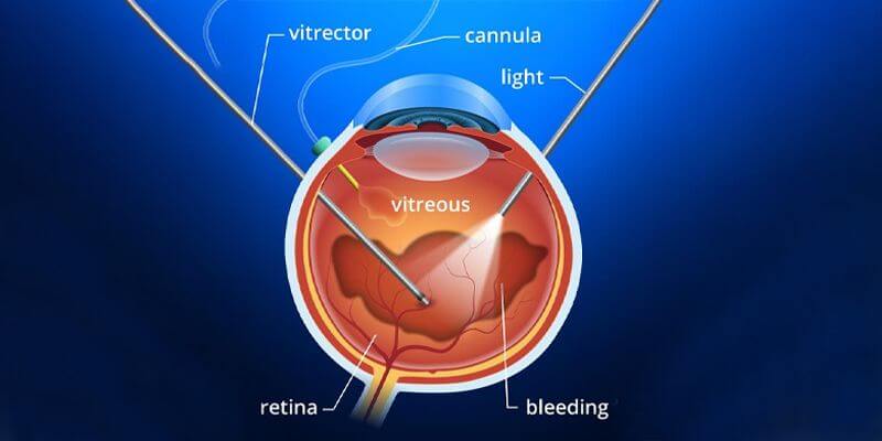
Introduction to Buckling Surgery Without Vitrectomy
Scleral buckling is a time-tested surgical technique used to repair rhegmatogenous retinal detachment. The operation works by indenting (or "buckling") the outer wall of the eye (the sclera) with a silicone element so that the retinal tear is brought into contact with the underlying pigment epithelium and choroid. The procedure may be performed as a local, segmental buckle (targeting one area) or as a circumferential/encircling buckle (around the globe), often combined with external cryotherapy or laser to seal the break. When performed without vitrectomy, the surgeon leaves the vitreous intact and uses buckle(s) plus subretinal fluid drainage or gas to encourage retinal reattachment.
Scleral buckling has become less commonly performed in some centres because modern vitrectomy techniques are versatile and easier to teach. Nevertheless, scleral buckling remains the preferred approach for many young, phakic patients and for certain tear locations or detachments where supporting the vitreous base externally is beneficial. Recent comparative studies continue to show that in properly selected cases buckling alone produces excellent anatomical success and, in some groups, better long-term visual outcomes and less cataract progression than PPV.
Causes and Risk Factors (Why a buckle may be chosen and relevant patient/disease factors)
Scleral buckle surgery is a time-tested treatment for rhegmatogenous retinal detachment (RRD)-a sight-threatening condition where a tear or hole in the retina allows fluid to separate it from the underlying retinal pigment epithelium. Although newer options like vitrectomy exist, a scleral buckle remains essential for certain patient and disease profiles.
Why retinal detachments happen (short background)
A rhegmatogenous retinal detachment occurs when a full-thickness retinal break allows liquefied vitreous to pass under the neurosensory retina, separating it from the retinal pigment epithelium. Common predisposing factors include posterior vitreous detachment (PVD), myopia (especially high myopia), lattice degeneration, trauma, prior ocular surgery, and certain inflammatory or degenerative retinal conditions.
Risk factors that affect the choice of scleral buckle (who is a better candidate)
Surgeons generally consider scleral buckling without vitrectomy when one or more of the following apply:
-
Phakic lens status (natural crystalline lens still present): buckling avoids entering the vitreous cavity and thus significantly reduces the risk of accelerating cataract formation compared with PPV. This is a major reason to favour buckling in younger, phakic patients.
-
Retinal breaks located anteriorly (at or near the vitreous base): these are often well treated by external indentation and cryotherapy.
-
Relatively recent (acute) detachments and fewer breaks: smaller extent detachments with limited subretinal fluid are often amenable to buckle repair.
-
Absence of dense vitreous hemorrhage, media opacity, or proliferative vitreoretinopathy (PVR): if visualization is poor or PVR is advanced, vitrectomy is often required.
Risk factors for failure or complications
Factors that increase the chance of recurrent detachment or complications after buckle-only surgery include extensive detachments, multiple or large posterior breaks, high myopia with posterior staphyloma, chronic longstanding detachments, preexisting PVR, and poor visualization of breaks preoperatively.
Symptoms and Signs (What patients present with and clinical findings)
Rhegmatogenous retinal detachment (RRD) - the main condition treated with a scleral buckle - results from a break, tear, or hole in the retina that allows vitreous fluid to separate the retinal layers. Patients typically present with sudden visual symptoms and distinctive clinical signs that guide urgent diagnosis and management.
Typical patient symptoms
Patients with a rhegmatogenous detachment commonly describe:
-
Sudden onset of floaters (new "specks" or cobwebs).
-
Flashes of light (photopsia), especially in peripheral vision.
-
A shadow/curtain spreading across part of the visual field (often peripheral first).
-
Blurred or decreased vision, which may progress if the macula becomes detached.
Timely presentation is critical - detachments involving the macula (central retina) carry a worse visual prognosis, and prompt repair offers the best chance of visual recovery.
Signs on clinical examination
Ophthalmic exam typically shows a detached, mobile neurosensory retina on indirect ophthalmoscopy, often with a visible retinal tear and subretinal fluid. Peripheral retinal tears may be localized with scleral depression. Optical coherence tomography (OCT) can document macular attachment status and help with prognostication. Clinical detection and accurate localization of all causative tears are essential for successful scleral buckle surgery.
Diagnosis (How the diagnosis and surgical plan are made)
Diagnosis and surgical planning for rhegmatogenous retinal detachment (RRD)-the condition treated by scleral buckle surgery-depend on detailed clinical examination, imaging, and precise mapping of retinal breaks. The goal is to confirm the diagnosis, identify the causative retinal tear(s), and choose the most effective surgical strategy (buckle type, placement, and whether vitrectomy is required).
-
Optical coherence tomography (OCT): to evaluate the macula and quantify foveal involvement.
-
Wide-field fundus photography / imaging: useful for documentation and surgical planning.
-
B-scan ultrasonography: invaluable when media opacity (dense cataract, vitreous hemorrhage) prevents direct visualization; helps detect the extent of detachment and excludes other posterior segment pathology.
Once a retinal detachment is confirmed, the retina specialist maps all retinal breaks, assesses the degree of subretinal fluid and PVR, and assesses lens status. Those findings determine whether a scleral buckle alone is a reasonable first choice or whether PPV (with or without buckle) is preferable. Modern practice increasingly individualizes the approach rather than following a single standard for all RRDs.
Treatment Options - focus on Buckling Without Vitrectomy
Scleral buckling without vitrectomy remains a well-established and highly effective surgical technique for repairing rhegmatogenous retinal detachment (RRD), especially in phakic, younger patients without a posterior vitreous detachment (PVD). This approach avoids entering the vitreous cavity, preserving the natural lens and minimizing intraocular complications.
Goals of treatment
-
Re-appose the retina to the retinal pigment epithelium, close retinal breaks, and limit further fluid ingress.
-
Restore and preserve as much vision as possible, minimize complications and the need for additional procedures.
Scleral buckling - procedural overview (buckle without vitrectomy)
-
Anesthesia: local block (peribulbar/retrobulbar) or general anesthesia depending on patient and surgeon preference.
-
Localization of breaks: intraoperative transillumination or preoperative localization guides buckle placement.
-
Cryopexy or external laser: applied to the retinal tear to produce adhesive chorioretinal scar.
-
Scleral buckle placement: silicone sponge or band is sutured to the sclera to indent the wall under the break (segmental or encircling).
-
Subretinal fluid drainage (optional): in some cases external drainage is performed to flatten the retina intraoperatively.
-
Closure and post-op positioning: if included, a gas tamponade may be used and patients are instructed on head positioning if needed.
Technical variations include the use of segmental buckles (targeted) versus encircling elements and whether to drain subretinal fluid. Surgeons choose the method based on break location, extent of detachment, and intraoperative findings.
When is buckle preferred vs vitrectomy?
-
Prefer buckle-alone: young, phakic patients with anterior breaks or uncomplicated detachments and good visualization. Buckling avoids intraocular instrumentation and the higher risk of postoperative cataract progression associated with vitrectomy.
-
Prefer PPV (or PPV ± buckle): pseudophakic eyes with posterior breaks, complex detachments with PVR or vitreous traction, dense vitreous hemorrhage, or media opacity. Combined procedures (PPV + buckle) may be used in selected complex cases.
Success rates and visual outcomes
Recent comparative studies and systematic reviews show similar primary anatomic success rates for scleral buckling and PPV in many RRD presentations when cases are appropriately selected. Some modern series report better final visual acuity and lower rates of cataract progression with buckling in phakic eyes compared with PPV. Despite the shift toward vitrectomy, the evidence supports the continued role of buckling in selected patients.
Postoperative care
-
Topical antibiotics and anti-inflammatories are prescribed.
-
Activity and positioning instructions depend on whether gas was used and the surgeon's preferences.
-
Follow-up is typically within 1 day, week 1, and then according to retinal reattachment progress.
Prevention and Management (how to prevent detachments and manage post-op course)
Prevention and postoperative management of retinal detachment-especially after scleral buckle surgery-focus on early detection of risk factors, protecting the retina from future tears, and supporting proper healing to maintain long-term visual stability.
Prevention of primary RRD
-
Regular retinal checks for high-risk patients (high myopes, lattice).
-
Prompt treatment of retinal tears or lattice degeneration with peripheral laser barricade or cryopexy in asymptomatic but high-risk lesions.
-
Protective eyewear for trauma prevention.
Strategies to reduce complications after buckle surgery
-
Careful patient selection (avoid buckle-alone where PVR or posterior tears are likely).
-
Meticulous surgical technique to minimize muscle trauma (reduces risk of strabismus) and to avoid scleral perforation.
-
Appropriate antisepsis to reduce infection risk and careful handling of explant materials.
Managing incomplete reattachment or recurrence
-
If reattachment fails or recurrent detachment occurs, options include revision of the buckle, buckle removal and PPV, or combined PPV with tamponade. Decision depends on cause of failure (e.g., missed posterior break, PVR). Modern practice accepts reoperation with PPV if necessary; outcomes are generally good when timely.
Complications of Buckling Surgery Without Vitrectomy
Scleral buckling without vitrectomy is a highly successful procedure for rhegmatogenous retinal detachment, but-as with any ocular surgery-it carries both intraoperative and postoperative risks. Most complications are rare, but awareness, prevention, and timely management are crucial to maintaining retinal reattachment and visual results.
Scleral buckling is generally safe but, like any surgery, carries risks. Important complications to know:
-
Induced myopia / refractive change: encircling elements commonly produce a myopic shift (often around −1 to −3 diopters depending on element and eye), which may be permanent.
-
Diplopia / strabismus: transient diplopia is common early after surgery; persistent strabismus occurs in a minority (rates reported from ~2-10% depending on series) and may require prism/glasses or strabismus surgery.
-
Buckle infection, extrusion, or erosion: infection is an uncommon but important cause of buckle removal (reported infection rates vary, commonly <5%). Explants (especially some older hydrogel materials) can swell or extrude long term and require removal.
-
Scleral thinning or perforation: rare but serious; care is taken while suturing.
-
Anterior segment ischemia: very rare but potentially vision-threatening-more likely if multiple rectus muscles are extensively manipulated.
-
Cystoid macular edema, epiretinal membrane formation, or progression to proliferative vitreoretinopathy (PVR): these can compromise visual recovery and may require further surgery.
If a buckle becomes symptomatic (pain, infection, persistent diplopia, extrusion), removal may be required; removal usually preserves retinal reattachment in most patients but is decided case-by-case.
Living with the Condition - patient expectations and rehabilitation
Living after scleral buckle surgery involves a recovery phase focused on protecting the eye, gradual visual rehabilitation, and adjusting to mild long-term changes in refraction or vision. With appropriate care and follow-up, most patients recover good functional vision and return to daily life within weeks to months.
Visual prognosis
-
Visual recovery depends primarily on macular status at the time of surgery (macula-on detachments do better), the duration of macular detachment, and preexisting ocular health. Prompt repair gives the best chance of good visual recovery. Some patients will not regain pre-detachment vision, particularly if the macula was detached for a long time or if PVR develops.
Practical advice for patients after a scleral buckle
-
Expect blurred vision initially and a period of adaptation to visual changes and possible induced myopia.
-
Avoid heavy lifting, straining, or activities that may increase intraocular pressure during early recovery (surgeon will specify restrictions).
-
If a gas bubble was used, avoid air travel until the gas is fully resorbed and follow surgeon instructions on timing - gas expansion at altitude can cause dangerous pressure rises.
-
Attend all follow-up visits - early detection of complications or recurrent detachment is key.
Long-term follow-up
-
Regular retinal examinations yearly or as advised, especially if the fellow eye is at risk.
-
Those with new symptoms (increased floaters, flashes, visual field shadowing) should seek immediate ophthalmic attention.
Top 10 Frequently Asked Questions about Buckling Surgery Without Vitrectomy
1. What is Buckling Surgery Without Vitrectomy?
Buckling surgery without vitrectomy is a specialized eye surgery used to
repair rhegmatogenous retinal detachment - a condition where the retina
separates from its underlying tissue due to a tear or hole.
Instead of removing the vitreous gel (as in a vitrectomy), the surgeon places a
silicone band or sponge (called a scleral buckle) around the white part
of the eye (the sclera). This buckle gently indents the eye wall, bringing it closer to
the detached retina and sealing the retinal tear.
The buckle acts as a permanent external support, allowing the retina to reattach naturally while preserving the eye's internal structure and vitreous gel.
This method is minimally invasive compared to vitrectomy and is especially useful in young or phakic patients (those with their natural lens intact), where preserving the vitreous is beneficial.
2. How Does Buckling Surgery Work Without Vitrectomy?
The surgery works on a simple mechanical principle: by indentation and
support.
When the retina detaches due to a tear, fluid seeps underneath it, separating it from
the underlying tissue. The scleral buckle pushes the outer wall of the eye inward,
helping the retinal tear close. At the same time, cryotherapy (freezing
treatment) is applied to the tear to create scar tissue that permanently
seals it.
Because the vitreous body isn't removed, the internal structure of the eye remains intact, and the eye's natural pressure dynamics are maintained. The fluid beneath the retina is either naturally absorbed over time or drained externally during surgery.
3. When is Buckling Surgery Recommended Over Vitrectomy?
Buckling surgery without vitrectomy is usually recommended in the following situations:
-
Uncomplicated retinal detachments caused by a single or small peripheral retinal break.
-
Younger patients, as their vitreous is more intact and less liquefied.
-
Phakic eyes, where avoiding vitrectomy helps prevent cataract development.
-
Cases without significant vitreous traction, meaning the vitreous gel isn't pulling on the retina.
-
Certain types of lattice degeneration or small round holes leading to detachment.
In contrast, vitrectomy is preferred for complex or giant retinal tears, posterior retinal breaks, or when there's dense vitreous hemorrhage obstructing visibility.
4. What Are the Steps Involved in the Surgery?
A typical scleral buckling procedure without vitrectomy involves these key steps:
-
Anesthesia: The eye is numbed using local or general anesthesia.
-
Exposure: The surgeon carefully exposes the sclera (white part of the eye).
-
Localization: Using indirect ophthalmoscopy, the retinal tear or detachment area is identified.
-
Cryopexy: The tear is sealed using cryotherapy, creating controlled scarring.
-
Placement of Buckle: A silicone band or sponge is sutured onto the sclera to indent the area under the tear.
-
Fluid Drainage (if needed): Subretinal fluid may be drained externally.
-
Closure: The conjunctiva is repositioned, and the eye is bandaged.
The surgery usually takes 60-90 minutes and is performed under sterile, microscopic conditions.
5. What Are the Benefits of Avoiding Vitrectomy in This Procedure?
Choosing buckling without vitrectomy offers several distinct benefits:
-
Preservation of the natural vitreous: Helps maintain eye structure and reduces internal complications.
-
Lower risk of cataract formation: Common after vitrectomy but minimal here.
-
Shorter recovery period: Less invasive and faster healing.
-
Reduced intraocular inflammation: As the vitreous remains untouched, there's less trauma.
-
Better for younger patients: Maintains the natural lens and ocular dynamics.
-
Fewer complications related to intraocular surgery, such as endophthalmitis or hemorrhage.
6. What Are the Possible Risks or Complications?
Although generally safe, buckling surgery without vitrectomy carries some potential risks, including:
-
Infection (endophthalmitis) - rare but serious.
-
Double vision (diplopia) due to eye muscle imbalance.
-
Discomfort or foreign body sensation from the buckle.
-
Exposure or migration of the buckle over time.
-
Re-detachment of the retina, requiring further surgery.
-
Refractive changes - the buckle can cause mild nearsightedness (myopia).
Most of these risks are manageable with timely follow-up and proper postoperative care.
7. How Successful is Buckling Surgery Without Vitrectomy?
The success rate of scleral buckling surgery without vitrectomy is
impressively high - around 85% to 90% for the first procedure.
If reoperation is required, overall success can reach over 95%.
Success largely depends on factors such as:
-
Early diagnosis and prompt treatment.
-
Size and location of the retinal tear.
-
Whether the macula (central retina) was detached.
-
The skill and experience of the surgeon.
When performed on suitable candidates, this surgery offers excellent long-term visual outcomes and stability.
8. What is the Recovery Process Like?
Postoperative recovery after buckling surgery without vitrectomy typically follows this timeline:
-
First 24-48 hours: Mild discomfort, redness, or swelling around the eye.
-
1-2 weeks: Vision starts to stabilize as the retina heals.
-
4-8 weeks: Most patients return to normal daily activities.
-
Full recovery: Around 6-8 weeks after surgery.
Patients are advised to:
-
Avoid strenuous activities, bending, or lifting.
-
Use prescribed antibiotic and steroid eye drops.
-
Sleep in a head position recommended by the surgeon.
-
Wear protective eyewear and avoid eye rubbing.
Regular follow-ups are crucial to monitor retinal stability and ensure the buckle remains in place.
9. Will My Vision Return to Normal After Surgery?
Visual recovery depends on how long the retina was detached and whether
the macula was involved.
If the macula was attached at the time of surgery, patients often regain near-normal
vision. If it was detached, some visual distortion or blurriness may remain, though
peripheral vision typically improves significantly.
Even when full visual restoration isn't possible, the main goal - preventing further vision loss and preserving functional sight - is usually achieved.
10. How Do I Prepare for and Care After the Surgery?
Preoperative preparation:
-
Undergo complete eye examination and imaging (OCT, ultrasound).
-
Discuss any medications or allergies with the doctor.
-
Avoid eating or drinking for 6-8 hours before surgery (if under general anesthesia).
Postoperative care:
-
Use prescribed medications as directed.
-
Keep the eye clean and avoid water entry for at least 2 weeks.
-
Attend all follow-up visits to track retinal healing.
-
Report any pain, sudden vision loss, or flashes immediately to your surgeon.
With good compliance, long-term prognosis after buckling surgery without vitrectomy is excellent.


