
Introduction to Canalicular Tear Repair
The eyes are among the most delicate and vital parts of the human body, not only essential for sight but also for expression, appearance, and emotional communication. Within this complex structure lies a small but crucial system called the lacrimal drainage system, responsible for draining tears from the eyes into the nose. The canaliculi (singular: canaliculus) are tiny channels within this system that carry tears from the puncta (tear openings) in the eyelids to the lacrimal sac.
A canalicular tear or canalicular laceration occurs when one of these fine channels is cut or damaged - most commonly due to trauma near the inner corner of the eyelid (the medial canthus). Such an injury can disrupt the tear drainage process, causing continuous tearing (epiphora), infection, or scarring if left untreated.
Canalicular tear repair is a delicate microsurgical procedure designed to restore the normal tear drainage pathway by reconnecting the severed canalicular ends. This is achieved using fine sutures and specialized stents that help keep the channel open while it heals. The surgery aims not only to preserve the aesthetic appearance of the eyelid but also to prevent long-term functional complications such as chronic tearing or recurrent infection.
Prompt repair of canalicular tears is critical - ideally within 24 to 48 hours after the injury. With modern surgical techniques and materials, the prognosis for successful restoration of tear drainage is excellent when performed by an experienced oculoplastic surgeon.
Causes and Risk Factors of Canalicular Tear
A canalicular tear (also called canalicular laceration) is a traumatic injury that affects the canaliculus - the small duct in the eyelid that drains tears from the eye into the nasal cavity. These injuries commonly occur near the inner corner of the eyelid and, if left untreated, can lead to chronic complications like excessive tearing (epiphora), scarring, and infection.
A. Common Causes
A canalicular tear is almost always the result of trauma to the eyelid or surrounding structures. The following are the most frequent causes:
-
Blunt or Sharp Eyelid Trauma:
The majority of canalicular injuries occur due to direct trauma to the inner corner of the eyelid. This may result from road accidents, sports injuries, falls, or physical altercations. -
Animal Bites:
Dog and cat bites are common causes, especially in children. Such injuries often involve deep tissue lacerations and carry a high risk of infection. -
Penetrating Foreign Objects:
Sharp objects such as sticks, glass shards, or metal fragments can cut through the eyelid, damaging the canalicular system. -
Post-Surgical or Iatrogenic Causes:
Rarely, the canaliculus may be inadvertently injured during eyelid or nasal surgeries if the anatomy is distorted or the injury is not recognized. -
Industrial or Work-Related Accidents:
Injuries caused by tools, machinery, or flying debris can also damage the delicate canalicular channels.
B. Risk Factors
Certain factors can worsen the extent of injury or make repair more complicated:
-
Delay in seeking treatment: Waiting too long after the injury allows tissue scarring, making repair difficult.
-
Associated injuries: Trauma that also affects the globe (eyeball) or facial bones complicates canalicular repair.
-
Infection: Particularly after animal bites, bacterial contamination can compromise healing.
-
Smoking and systemic illness: Poor circulation or chronic conditions like diabetes can delay wound healing.
-
Tissue loss or avulsion: When large areas of eyelid tissue are missing, microsurgical reconstruction is more complex.
Timely and accurate diagnosis followed by immediate surgical intervention are key factors that determine the success of canalicular tear repair.
Symptoms and Signs of Canalicular Tear
A canalicular tear is usually apparent following trauma, but the extent of the damage may not always be obvious without a detailed examination. Recognizing the signs and symptoms early ensures that the injury is properly treated before complications arise.
A. Common Symptoms
-
Excessive tearing (epiphora): One of the earliest and most common symptoms, as the tears are unable to drain normally.
-
Swelling and bruising: The eyelid near the inner corner becomes swollen or discolored following trauma.
-
Pain or tenderness: The affected area may be sore to touch, especially shortly after injury.
-
Discharge or crusting: If infection develops, the patient may notice pus or crust formation around the eye.
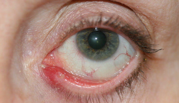
B. Visible Signs Noted by Clinicians
-
Laceration near the inner canthus: A cut or wound close to the tear duct area often indicates canalicular involvement.
-
Displacement of the punctum: The small tear opening may appear irregular, widened, or out of position.
-
Gap in the eyelid margin: If the eyelid has been torn, a visible gap may be present.
-
Bleeding or exposed tissue: The canalicular ends may occasionally be visible in deep lacerations.
C. Functional Signs
-
Persistent tearing even after the wound heals
-
Recurrent eye infections or conjunctivitis
-
Decreased ability of tears to drain properly, causing constant wetness of the lower eyelid
Patients who sustain any eyelid injury near the tear duct should always seek immediate evaluation by an ophthalmologist or oculoplastic specialist to rule out canalicular damage.
Diagnosis of Canalicular Tear
The diagnosis of a canalicular tear (also called canalicular laceration) relies on a detailed clinical evaluation supported by specialized ophthalmic techniques. The goal is to identify the site and extent of damage to the eyelid's tear drainage ducts while ensuring no associated ocular or facial trauma is overlooked.
A. Medical History
Diagnosis begins with a thorough medical history, including details about how the injury occurred, timing, the presence of animal bites or foreign bodies, and any previous eye surgeries. Tetanus status and history of systemic illnesses are also reviewed.
B. Clinical Examination
The doctor performs a meticulous examination using magnification:
-
Inspection of the eyelid margins for lacerations or displacement of the punctum.
-
Gentle probing or irrigation of the tear drainage system to check for obstruction.
-
Use of fluorescein dye to help identify the cut ends of the canaliculus.
-
Examination of the eye itself to ensure there is no globe rupture, corneal damage, or intraocular injury.
C. Imaging Studies
While most canalicular injuries are diagnosed clinically, imaging such as CT scans may be ordered in cases of facial fractures or suspected foreign bodies. MRI may be used for soft tissue assessment in complex or delayed cases.
D. Classification
-
Monocanalicular Tear: Involves only one canaliculus (upper or lower).
-
Bicanalicular Tear: Involves both upper and lower canaliculi, typically in severe trauma.
This classification helps determine the complexity of repair and the type of stent or surgical technique required.
Treatment Options for Canalicular Tear Repair
Canalicular tear repair is almost always a surgical procedure. The main goal is to restore the anatomy and function of the lacrimal drainage system so that tears can flow normally again.
A. Timing of Repair
Ideally, the repair should be performed within 24 to 48 hours of injury to prevent scarring, infection, and tissue contraction. Delayed repair is possible but becomes more technically challenging and less predictable.
B. Surgical Techniques
-
Microsurgical Realignment:
Using an operating microscope, the surgeon identifies and reconnects the two ends of the torn canaliculus. Fine sutures are used to close the canalicular tissue. -
Stenting (Intubation):
A small silicone stent or tube is placed through the canaliculus and into the lacrimal drainage system to maintain its shape during healing.-
Monocanalicular Stent: Used when only one canaliculus is injured.
-
Bicanalicular Stent: Used when both canaliculi are involved.
The stent is usually left in place for 6 to 12 weeks and then removed in the clinic.
-
-
Repair of Overlying Eyelid:
Once the canalicular system is reconnected, the surrounding eyelid tissue is carefully sutured to restore appearance and function. -
Antibiotics and Wound Care:
Topical and oral antibiotics are given, particularly in animal bite cases, to prevent infection. Pain relief and anti-inflammatory medication are also prescribed.
C. Alternative or Adjunct Treatments
In minor injuries or when repair is delayed, observation or secondary reconstruction may be considered. However, direct repair remains the gold standard for functional success.
Prevention and Management of Canalicular Tear Repair
The prevention and management of canalicular tear repair aim to protect the lacrimal drainage system, restore normal tear flow, and avoid future complications such as epiphora (tear overflow) or canalicular obstruction. Successful outcomes rely on prompt surgical intervention, proper stenting techniques, postoperative care, and patient compliance.
A. Prevention of Canalicular Tears
-
Protective Eyewear: Use in high-risk environments such as construction, laboratories, or while cycling.
-
Safe Handling of Pets: Educate children and adults to avoid rough play with animals to prevent bites.
-
Caution in Sports and Outdoor Activities: Helmets and face guards reduce trauma risk.
-
Surgical Awareness: Surgeons operating near the eyelid or medial canthus must remain cautious to avoid accidental canalicular injury.
B. Postoperative Management
-
Eye Protection: Wear protective eyewear or shields, especially during sleep.
-
Medication Adherence: Use antibiotic drops or ointments as directed.
-
Avoid Rubbing the Eye: This can dislodge the stent or disrupt the sutures.
-
Regular Follow-Up: Monitor for signs of infection, redness, pain, or excessive tearing.
-
Stent Removal: Usually performed 6-12 weeks post-surgery when healing is complete.
A well-followed postoperative plan ensures successful outcomes and reduces the risk of canalicular blockage or infection.
Complications of Canalicular Tear Repair
While modern microsurgical techniques make canalicular tear repair highly successful, complications can still occur. These may be early (during healing) or late (months to years after surgery).
A. Early Complications
-
Infection: Especially common in animal bites or contaminated wounds.
-
Bleeding or Hematoma Formation: Small bruises or blood clots under the skin may develop.
-
Stent Displacement: The silicone tube can shift or come out prematurely.
-
Eyelid Malposition: Improper healing may cause lid notching or distortion near the inner canthus.
B. Late Complications
-
Canalicular Stenosis (Narrowing): Scar tissue may partially close the repaired canaliculus.
-
Obstruction or Functional Failure: Persistent tearing despite anatomically successful repair.
-
Visible Scarring: Small scars may form along the eyelid margin, although these usually fade with time.
-
Need for Revision Surgery: In rare cases, a secondary procedure may be required to re-open the canaliculus or adjust the stent.
Proper surgical technique, timely repair, and attentive follow-up can minimize these risks and improve the likelihood of full recovery.
Living with the Condition After Canalicular Tear Repair
After successful canalicular repair, most patients regain normal tear drainage and a natural appearance of the eyelid. Healing is generally smooth, and long-term outcomes are excellent when postoperative care is followed.
A. What to Expect After Surgery
-
Mild swelling and bruising for 1-2 weeks.
-
Stent visibility - a small portion may be seen at the corner of the eye, which is normal.
-
Some tearing is expected initially and usually subsides as healing completes.
-
Normal vision and eyelid function return within a few weeks.
B. Long-Term Care
-
Attend all follow-up visits to ensure the stent remains in position.
-
Maintain good eye hygiene to prevent infections.
-
Report any recurrent tearing, redness, or discharge promptly.
-
Once healed, most patients can return to all daily activities, sports, and work without restrictions.
C. Emotional and Cosmetic Recovery
Beyond physical healing, canalicular repair restores self-confidence and comfort, especially for patients concerned about appearance after trauma. The surgery offers both functional and aesthetic benefits, helping patients regain normal tear drainage and eyelid contour.
Top 10 Frequently Asked Questions about Canalicular Tear Repair
1. What is Canalicular Tear Repair?
Canalicular tear repair is a microsurgical procedure used to restore the normal function of the tear drainage system when the canaliculus (the small duct in the eyelid that drains tears) is damaged or torn.
This damage is often caused by trauma, such as a facial injury, dog bite, or sharp object. The surgery involves reconnecting the torn canaliculus and often inserting a tiny silicone stent or tube to keep the tear duct open during healing.
The goal of this procedure is to prevent chronic tearing (epiphora) and maintain proper tear flow from the eyes to the nasal cavity.
2. What Causes Canalicular Tears or Damage?
Canalicular injuries are commonly caused by:
-
Blunt or penetrating trauma to the face (sports injuries, car accidents, falls).
-
Animal bites, especially dog bites.
-
Lacerations near the eyelid margin caused by sharp objects or surgery.
-
Infections or chronic inflammation of the tear ducts.
-
Tumors or cysts affecting the lacrimal drainage pathway.
Because the canaliculus is very small and fragile, even minor trauma near the inner corner of the eye can result in a tear.
3. How Do I Know If I Have a Canalicular Tear?
Common symptoms of a canalicular tear include:
-
Persistent tearing or watery eyes (epiphora).
-
Swelling or tenderness near the inner corner of the eyelid.
-
Visible cut or laceration near the tear duct area.
-
Discharge or infection around the eye.
-
Difficulty draining tears, causing tears to overflow onto the cheeks.
If you experience these symptoms after a facial injury, it's crucial to seek prompt medical attention from an ophthalmologist or oculoplastic surgeon.
4. When is Canalicular Tear Repair Necessary?
Immediate surgical repair is recommended when:
-
There is a laceration involving the eyelid near the tear duct.
-
You experience persistent tearing or a blocked tear drainage pathway.
-
The injury disrupts the normal tear drainage anatomy.
Prompt repair - ideally within 24 to 48 hours of the injury - provides the best chance of restoring normal tear function and preventing long-term complications such as scarring or duct obstruction.
5. How is Canalicular Tear Repair Performed?
The procedure is typically done under local anesthesia with sedation or general anesthesia. The steps usually include:
-
Cleaning and preparing the wound area.
-
Using a microscope to locate the torn ends of the canaliculus.
-
Reconnecting the tear duct using fine sutures or a stent (a small silicone tube).
-
The stent helps keep the canal open during healing and is usually removed after 6-12 weeks.
-
The skin and eyelid are carefully closed to restore appearance and function.
The surgery usually takes 30-60 minutes depending on the complexity of the tear.
6. What is the Recovery Process After Canalicular Tear Repair?
Recovery is generally smooth when post-operative instructions are followed. Here's what to expect:
-
Mild swelling, bruising, or discomfort around the eye for 1-2 weeks.
-
Use of prescribed antibiotic and anti-inflammatory eye drops.
-
Avoid rubbing or pressing on the eye.
-
Stitches may dissolve on their own or be removed in a follow-up visit.
-
The silicone stent is typically removed after 6-12 weeks once healing is complete.
Most patients resume normal daily activities within a few days to a week, but strenuous exercise should be avoided during early recovery.
7. Are There Any Risks or Complications Associated with Canalicular Tear Repair?
As with any surgery, there are potential risks, though they are rare when performed by an experienced oculoplastic surgeon. Possible complications include:
-
Infection or inflammation at the surgical site.
-
Scarring or blockage of the canaliculus.
-
Displacement or premature loss of the stent.
-
Incomplete tear drainage, leading to continued tearing.
-
Eyelid malposition or cosmetic irregularities.
Timely follow-up appointments and proper care greatly reduce the chances of complications.
8. What Happens If a Canalicular Tear Is Not Repaired?
If left untreated, a canalicular tear can lead to chronic tearing, infection, and scarring of the tear drainage pathway. The tear duct may become permanently blocked, requiring more complex surgery later (such as dacryocystorhinostomy, or DCR).
Prompt surgical repair not only restores function but also helps achieve better cosmetic and long-term results.
9. Is Canalicular Tear Repair Painful?
The surgery itself is painless, as it is performed under local or
general anesthesia.
After the procedure, patients may experience mild soreness or swelling
for a few days, which can be managed with prescribed pain medication and cold
compresses.
Most people describe the post-surgery discomfort as minimal and short-lived.
10. How Successful Is Canalicular Tear Repair?
When performed by a skilled ophthalmic or oculoplastic surgeon, canalicular tear repair has a very high success rate - typically 85-95%.
The use of silicone stents helps maintain the canal's shape and function during healing, reducing the risk of blockage. Most patients experience normal tear drainage and minimal scarring after complete recovery.
