
Introduction to Endoscopic Discectomy
Endoscopic Discectomy is one of the most remarkable advancements in modern spinal surgery, offering patients an effective and minimally invasive solution to relieve chronic back pain, leg pain (sciatica), and nerve compression caused by herniated or degenerated spinal discs.
Unlike traditional open spine surgery, which involves large incisions, extensive muscle dissection, and long recovery periods, endoscopic discectomy allows surgeons to access the affected spinal disc through a tiny incision (as small as 8 mm) using an endoscope - a thin tube equipped with a high-resolution camera and surgical instruments.
This advanced surgical technique enables direct visualization of the damaged disc and compressed nerve roots, allowing surgeons to precisely remove only the herniated or protruding portion of the disc while preserving surrounding muscles, ligaments, and bone. The result is less postoperative pain, minimal blood loss, shorter hospital stay, and quicker recovery compared to traditional spine surgery.
In a healthy spine, the intervertebral discs act as shock absorbers, cushioning the vertebrae and facilitating smooth movement. When one of these discs ruptures or bulges due to aging, injury, or degeneration, the inner gel-like material (nucleus pulposus) can press on nearby spinal nerves, causing sharp pain, numbness, or weakness along the back, buttocks, or legs.
Endoscopic Discectomy effectively removes this herniated tissue through a minimally invasive approach, restoring the natural anatomy and relieving nerve pressure. Today, this surgery is used to treat conditions such as lumbar disc herniation, cervical disc herniation, spinal stenosis, foraminal narrowing, and sciatica.
By 2025, endoscopic discectomy has become a gold-standard, globally accepted spinal procedure, recognized for its precision, safety, and ability to return patients to their normal lives faster than ever before.
Causes and Risk Factors Leading to Endoscopic Discectomy
The primary reason for undergoing endoscopic discectomy is disc herniation - a condition in which the soft inner gel of a spinal disc pushes out through its tougher outer layer and irritates the surrounding spinal nerves. This can occur in the cervical (neck), thoracic (mid-back), or lumbar (lower back) regions, though lumbar herniation is the most common.
A. Common Causes
-
Degenerative Disc Disease (DDD):
As people age, spinal discs naturally lose water content, flexibility, and strength, making them prone to tearing and bulging. -
Injury or Trauma:
A sudden impact, heavy lifting, or twisting motion can cause the disc to rupture or shift, compressing the spinal nerves. -
Repetitive Strain or Overuse:
Occupations involving frequent bending, lifting, or vibration (such as driving or construction work) increase the risk of disc injury. -
Poor Posture:
Chronic slouching or sitting in awkward positions puts uneven pressure on spinal discs. -
Obesity:
Excess body weight exerts continuous stress on the spine, accelerating disc degeneration. -
Genetic Factors:
Some individuals inherit a predisposition to weak discs and early degeneration. -
Smoking:
Nicotine reduces blood flow to the spine, impairing the healing and regeneration of disc tissue. -
Lack of Exercise:
Weak core and back muscles lead to instability and increased stress on spinal discs.
B. Risk Factors
Certain individuals are more prone to disc herniation and may eventually require surgical intervention:
-
Age: Most common between ages 30-60.
-
Occupational Hazards: Jobs requiring repetitive lifting or bending.
-
Sedentary Lifestyle: Sitting for long hours, especially without back support.
-
Sports Injuries: Especially in weightlifting, gymnastics, or contact sports.
-
Previous Back Injuries: Weakens spinal structures over time.
When the herniated disc begins to compress a nerve root or the spinal cord, symptoms become chronic and disabling - making Endoscopic Discectomy an appropriate and often life-changing treatment option.
Symptoms and Signs Indicating the Need for Endoscopic Discectomy
The symptoms of a herniated or degenerated disc depend on the location and severity of the nerve compression. The most commonly affected area is the lumbar spine, which supports much of the body's weight.
A. Common Symptoms
-
Severe Lower Back Pain:
Sharp, aching, or burning pain that worsens with movement, coughing, or sitting. -
Radiating Leg Pain (Sciatica):
Pain traveling down the buttock, thigh, calf, or foot - caused by compression of the sciatic nerve. -
Numbness or Tingling:
Sensations of “pins and needles” in the legs or feet, indicating nerve irritation. -
Muscle Weakness:
Difficulty walking, lifting objects, or standing due to weakened muscles in the affected limb. -
Limited Range of Motion:
Stiffness and reduced flexibility in the spine. -
Pain that Worsens When Sitting:
Sitting increases disc pressure and nerve compression. -
Neck or Shoulder Pain (Cervical Discs):
In cervical herniations, pain radiates into the arms and hands.
B. Advanced or Emergency Symptoms
-
Loss of Bladder or Bowel Control (Cauda Equina Syndrome): Requires immediate surgery.
-
Progressive Weakness or Numbness: Sign of worsening nerve damage.
-
Inability to Stand or Walk Properly: Indicates severe nerve root compression.
These symptoms can severely affect a person's daily life, limiting movement, sleep, and work performance. When pain persists for more than 6-8 weeks despite medication and therapy, Endoscopic Discectomy becomes a medically recommended solution.
Diagnosis and Evaluation Before Endoscopic Discectomy
Before performing endoscopic discectomy, a thorough evaluation is necessary to confirm that the pain originates from a herniated disc and that surgery is the most appropriate treatment.
A. Clinical Evaluation
-
Medical History:
The surgeon reviews pain onset, duration, prior injuries, and treatment attempts. -
Physical Examination:
Includes posture analysis, palpation for tenderness, range-of-motion testing, and nerve reflex checks. -
Neurological Assessment:
Tests muscle strength, sensory response, and reflexes to pinpoint the affected nerve.
B. Imaging Tests
-
Magnetic Resonance Imaging (MRI):
The most accurate method to visualize disc herniation and nerve compression. -
CT Scan:
Provides detailed images of spinal bones and disc protrusions. -
X-rays:
Shows spinal alignment, bone spurs, or degenerative changes. -
Myelogram:
A dye-based imaging technique used when MRI is contraindicated. -
Electromyography (EMG):
Measures electrical activity of muscles to detect nerve damage.
C. Indications for Surgery
-
Pain lasting over 6-12 weeks despite conservative therapy.
-
Progressive leg weakness or numbness.
-
MRI-confirmed disc herniation compressing the nerve root.
-
Disabling pain interfering with work or daily life.
Once confirmed, the patient is prepared for Endoscopic Discectomy, which offers targeted relief with minimal disruption to normal anatomy.
Treatment Options and The Endoscopic Discectomy Procedure
A. Non-Surgical Treatments
Prior to surgery, patients are usually managed with:
-
Pain medications (NSAIDs, muscle relaxants).
-
Physiotherapy and stretching exercises.
-
Epidural steroid injections to reduce inflammation.
-
Lifestyle modifications like ergonomic adjustments and rest.
If these do not provide lasting relief, surgical intervention becomes the best course of action.
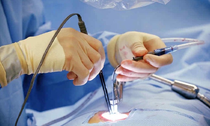
B. Endoscopic Discectomy Procedure (Step-by-Step)
1. Anesthesia and Positioning
The surgery is performed under local, spinal, or general anesthesia, depending on the patient's condition. The patient is positioned either on their side or face down on a radiolucent table.
2. Incision and Access
A small incision (less than 1 cm) is made over the affected disc area. Using X-ray or fluoroscopic guidance, a working cannula (hollow tube) is inserted to reach the herniated disc.
3. Insertion of the Endoscope
Through the cannula, an endoscope equipped with a micro-camera, light, and surgical tools is inserted. The surgeon can visualize the disc, nerve, and surrounding tissues on a monitor in real-time.
4. Disc Removal
The herniated or extruded disc fragment pressing on the nerve is carefully removed using micro-forceps or laser assistance. The remaining disc is sculpted to relieve compression.
5. Closure
After ensuring the nerve is fully decompressed, instruments are withdrawn, and the small incision is closed with a single stitch or skin adhesive.
6. Duration and Recovery
-
Surgery Time: 45-90 minutes.
-
Hospital Stay: Same-day discharge or 24-hour observation.
-
Mobility: Patients can walk within a few hours post-surgery.
Prevention and Postoperative Management
A. Prevention
Though disc herniation cannot always be avoided, risk reduction is possible through:
-
Regular core-strengthening exercises.
-
Maintaining a healthy body weight.
-
Avoiding prolonged sitting or poor posture.
-
Using proper lifting techniques (bend knees, not back).
-
Staying hydrated and avoiding smoking.
B. Postoperative Care
-
Immediate Recovery:
Pain is minimal and managed with mild analgesics. -
Activity Level:
Patients are encouraged to walk within hours; avoid strenuous activities for 4-6 weeks. -
Physiotherapy:
Begins within a week to strengthen muscles and restore flexibility. -
Wound Care:
Keep incision clean and dry; follow-up for suture removal if required. -
Return to Work:
Light work in 1-2 weeks, full activity in 6-8 weeks.
With proper rehabilitation, most patients experience complete recovery within 2-3 months and can resume normal, pain-free lives.
Complications of Endoscopic Discectomy
Endoscopic Discectomy is among the safest spinal procedures, but as with any surgery, complications may occur in rare cases.
A. Common Minor Complications
-
Mild swelling or soreness at incision site.
-
Temporary leg numbness or tingling.
-
Muscle spasms.
-
Minimal blood loss or bruising.
B. Rare Serious Complications
-
Infection or abscess formation (prevented with antibiotics).
-
Dural tear leading to cerebrospinal fluid leakage.
-
Recurrent disc herniation (5-10% of cases).
-
Nerve injury or chronic pain (very rare).
With skilled surgeons and advanced endoscopic technology, complication rates remain below 3-5%, significantly lower than in traditional open spine surgery.
Living After Endoscopic Discectomy
A. Recovery and Rehabilitation
After surgery, most patients experience immediate relief from leg pain and can return to normal walking within 24 hours. A customized rehabilitation plan focuses on:
-
Restoring flexibility and strength.
-
Maintaining correct posture.
-
Avoiding repetitive bending or twisting.
-
Long-term physical therapy for back health.
B. Long-Term Lifestyle Adjustments
-
Continue core-strengthening exercises like yoga or Pilates.
-
Avoid smoking and heavy lifting.
-
Maintain proper ergonomics at work.
-
Regular check-ups for spinal wellness.
C. Emotional and Physical Outcome
Patients report:
-
80-95% improvement in pain and mobility.
-
Better sleep and quality of life.
-
Minimal scarring and cosmetic satisfaction.
Endoscopic Discectomy gives individuals a second chance at living pain-free - regaining the freedom to move, work, and enjoy life fully again.
Top 10 Frequently Asked Questions about Endoscopic Discectomy
1. What is Endoscopic Discectomy?
Endoscopic Discectomy is a minimally invasive spine surgery performed to remove a herniated or slipped disc that is pressing on a spinal nerve. The surgeon uses an endoscope (a small camera) inserted through a tiny incision to visualize the disc and surrounding tissues. Special surgical instruments are then used to remove the damaged portion of the disc, relieving pain and nerve compression while preserving most of the healthy disc structure.
2. Who needs Endoscopic Discectomy?
Endoscopic discectomy is recommended for patients suffering from:
-
Lumbar or cervical disc herniation
-
Sciatica (leg pain due to nerve compression)
-
Chronic lower back or neck pain unresponsive to medication or therapy
-
Numbness or weakness in arms or legs caused by disc pressure
It is typically advised when conservative treatments like physiotherapy, rest, or injections fail to provide relief after 6-8 weeks.
3. How is Endoscopic Discectomy different from traditional spine surgery?
Unlike open spine surgery, Endoscopic Discectomy requires:
-
A tiny incision (about 8-10 mm)
-
No muscle cutting or bone removal
-
Minimal blood loss and tissue damage
-
Faster recovery and less postoperative pain
Patients often go home the same day and return to normal activities much sooner than after open discectomy or laminectomy.
4. What happens during the Endoscopic Discectomy procedure?
During the procedure:
-
The patient is given local or general anesthesia.
-
A small incision is made near the affected spine area.
-
The surgeon inserts an endoscope to locate the herniated disc.
-
Using specialized tools, the damaged disc fragments are carefully removed.
-
The incision is closed with one or two stitches or adhesive strips.
The entire surgery usually takes 30-60 minutes, depending on the location and severity of the disc herniation.
5. Is Endoscopic Discectomy painful?
The procedure itself is not painful since anesthesia is used. Postoperatively, patients may experience mild soreness or discomfort at the incision site, which usually subsides within a few days. Pain is significantly less than with traditional open spine surgery.
6. What are the benefits of Endoscopic Discectomy?
Key advantages of this procedure include:
-
Minimally invasive approach with tiny incision
-
Short hospital stay or same-day discharge
-
Faster recovery and less postoperative pain
-
Minimal scar and tissue trauma
-
Early return to work and normal activities
-
High success rate (over 90%) in relieving leg and back pain
7. What are the possible risks or complications?
Although rare, potential risks include:
-
Infection at the incision site
-
Nerve root injury
-
Recurrent disc herniation
-
Spinal fluid leak (dural tear)
-
Temporary numbness or tingling
Choosing an experienced spine surgeon significantly reduces the risk of complications.
8. How long is the recovery after Endoscopic Discectomy?
Recovery is typically quick:
-
Hospital stay: Same day or overnight
-
Return to light activities: Within 1-2 weeks
-
Resume normal work: In 2-4 weeks
-
Full recovery: Around 6 weeks
Patients are advised to follow a structured physiotherapy program to strengthen back muscles and prevent recurrence.
9. How successful is Endoscopic Discectomy?
Endoscopic discectomy has a success rate of 90-95% in relieving leg or arm pain caused by disc herniation. Patients experience significant pain reduction, improved mobility, and enhanced quality of life. However, maintaining proper posture, regular exercise, and weight control is essential for long-term success.
10. What precautions should I take after Endoscopic Discectomy?
After surgery, you should:
-
Avoid heavy lifting or twisting for at least 4-6 weeks
-
Follow your doctor's rehabilitation plan
-
Maintain good posture while sitting and standing
-
Do back-strengthening exercises as advised
-
Keep the incision area clean and dry until healed
Following these precautions ensures a smooth recovery and minimizes the risk of re-injury.
