
Introduction to Mitral Valve Repair
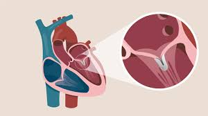
The human heart is a remarkably efficient organ made up of four chambers and four valves that work together to circulate blood throughout the body. Among these, the mitral valve is one of the most important. It is located between the left atrium and the left ventricle and functions as a one-way gate that allows oxygen-rich blood to flow from the atrium to the ventricle. When the valve becomes damaged, thickened, or too loose, it fails to close or open properly, disrupting blood flow. This condition is known as mitral valve disease.
Mitral valve repair is a surgical or minimally invasive procedure designed to restore the valve's normal function without replacing it entirely. During the procedure, surgeons reshape, tighten, or reinforce the existing valve tissue to eliminate leakage (regurgitation) or improve blood flow (in cases of stenosis). Compared to valve replacement, repair is preferred in most cases because it preserves the patient's natural valve, maintains normal heart function, and has a lower risk of complications such as infection or blood clots.
Advancements in cardiac surgery now allow many patients to undergo minimally invasive or robotic-assisted mitral valve repair, which leads to faster recovery, smaller scars, and shorter hospital stays. Mitral valve repair is not just a treatment—it's a life-saving procedure that restores quality of life, extends survival, and enables patients to return to normal daily activities with improved heart performance.
Causes and Risks of Mitral Valve Disease (Leading to Repair)
Mitral valve repair is typically performed to correct mitral regurgitation or mitral stenosis.
-
Mitral Regurgitation (MR) occurs when the valve does not close properly, causing blood to leak backward into the left atrium.
-
Mitral Stenosis (MS) happens when the valve becomes narrowed or stiff, limiting the amount of blood flowing from the atrium to the ventricle.
The underlying causes vary widely:
-
Degenerative valve disease: The most common cause in
developed countries. It occurs due to age-related changes or genetic
factors that weaken the valve tissues, leading to prolapse (floppy valve
leaflets).
-
Rheumatic heart disease: Still prevalent in developing
regions, including parts of Asia and Africa. It results from untreated
streptococcal infections that cause inflammation and scarring of the
valve.
-
Ischemic heart disease: A heart attack or coronary
artery disease may damage the structures supporting the valve, causing
it to leak.
-
Infective endocarditis: Bacterial infection of the heart
lining can destroy valve tissue, leading to severe leakage.
-
Congenital abnormalities: Some individuals are born with
malformed valve leaflets or supporting structures.
-
Connective tissue disorders: Conditions such as Marfan
syndrome can weaken the valve's elastic fibers.
Risk factors that increase the likelihood of mitral valve disease include
advanced age, previous rheumatic fever, uncontrolled hypertension, obesity, high
cholesterol, and smoking. Left untreated, these conditions can lead to
progressive valve damage, heart enlargement, pulmonary hypertension, and
ultimately heart failure.
Degenerative valve disease: The most common cause in developed countries. It occurs due to age-related changes or genetic factors that weaken the valve tissues, leading to prolapse (floppy valve leaflets).
Rheumatic heart disease: Still prevalent in developing regions, including parts of Asia and Africa. It results from untreated streptococcal infections that cause inflammation and scarring of the valve.
Ischemic heart disease: A heart attack or coronary artery disease may damage the structures supporting the valve, causing it to leak.
Infective endocarditis: Bacterial infection of the heart lining can destroy valve tissue, leading to severe leakage.
Congenital abnormalities: Some individuals are born with malformed valve leaflets or supporting structures.
Connective tissue disorders: Conditions such as Marfan syndrome can weaken the valve's elastic fibers.
Symptoms and Signs of Mitral Valve Disease
Mitral valve disease can remain silent for years before producing noticeable symptoms. As the valve deteriorates, the heart must work harder to maintain normal circulation, leading to physical signs that should never be ignored.
Common symptoms include:
-
Shortness of breath, particularly during exertion or when lying flat.
-
Fatigue and weakness due to reduced cardiac output.
-
Heart palpitations, often due to atrial fibrillation caused by left
atrial enlargement.
-
Chest discomfort or tightness, especially with exertion.
-
Swelling in the ankles, feet, or abdomen due to fluid buildup.
-
Persistent cough, sometimes producing frothy sputum due to fluid in the
lungs.
Physical examination by a doctor may reveal:
-
A distinct heart murmur heard through a stethoscope.
-
Irregular heartbeat (arrhythmia).
-
Signs of heart failure such as fluid retention or lung congestion.
Shortness of breath, particularly during exertion or when lying flat.
Fatigue and weakness due to reduced cardiac output.
Heart palpitations, often due to atrial fibrillation caused by left atrial enlargement.
Chest discomfort or tightness, especially with exertion.
Swelling in the ankles, feet, or abdomen due to fluid buildup.
Persistent cough, sometimes producing frothy sputum due to fluid in the lungs.
-
A distinct heart murmur heard through a stethoscope.
-
Irregular heartbeat (arrhythmia).
-
Signs of heart failure such as fluid retention or lung congestion.
If left untreated, the symptoms progressively worsen. Early identification and treatment of valve disease significantly improve outcomes, preventing irreversible heart muscle damage.
Diagnosis of Mitral Valve Disease
Diagnosing mitral valve disease requires a combination of clinical evaluation, imaging, and sometimes invasive testing. The goal is to accurately assess the valve's structure, the degree of leakage or narrowing, and its impact on heart function.
Key diagnostic methods include:
-
Echocardiogram (TTE or TEE): This ultrasound-based test
is the gold standard for assessing valve anatomy and function. It shows
how well the valve opens and closes and whether there is any
regurgitation or stenosis.
-
Electrocardiogram (ECG): Detects rhythm abnormalities
like atrial fibrillation.
-
Chest X-ray: Helps identify heart enlargement and fluid
buildup in the lungs.
-
Cardiac MRI or CT scan: Provides detailed 3D images of
the valve and surrounding heart tissue.
-
Cardiac catheterization: Measures pressures inside the
heart and may be used to evaluate for coexisting coronary artery disease
before surgery.
Echocardiogram (TTE or TEE): This ultrasound-based test is the gold standard for assessing valve anatomy and function. It shows how well the valve opens and closes and whether there is any regurgitation or stenosis.
Electrocardiogram (ECG): Detects rhythm abnormalities like atrial fibrillation.
Chest X-ray: Helps identify heart enlargement and fluid buildup in the lungs.
Cardiac MRI or CT scan: Provides detailed 3D images of the valve and surrounding heart tissue.
Cardiac catheterization: Measures pressures inside the heart and may be used to evaluate for coexisting coronary artery disease before surgery.
Once diagnosed, the heart team evaluates whether the valve can be repaired or needs to be replaced. Repair is the preferred option when the valve tissue is flexible and structurally salvageable. Patients with degenerative or prolapse-related regurgitation usually have the best outcomes with repair.
Treatment Options: Surgical and Minimally Invasive Repair
Mitral valve repair focuses on restoring the valve's natural function rather than replacing it with a mechanical or biological prosthesis. The procedure can be done through open-heart surgery or minimally invasive techniques, depending on the patient's condition.
Common surgical repair techniques include:
-
Annuloplasty: The surgeon places a supportive ring or
band around the valve to reshape and tighten the annulus (the valve's
base). This ensures the leaflets close properly.
-
Leaflet repair: Damaged or floppy valve leaflets are
reshaped, trimmed, or repositioned to restore their coaptation.
-
Chordal replacement: Artificial chords (made of durable
material like Gore-Tex) replace ruptured or stretched chordae tendineae,
which anchor the valve leaflets.
-
Commissurotomy: In mitral stenosis, fused valve leaflets
are separated to improve blood flow.
-
Patch reconstruction: Damaged areas may be rebuilt with
tissue patches to restore valve structure.
Annuloplasty: The surgeon places a supportive ring or band around the valve to reshape and tighten the annulus (the valve's base). This ensures the leaflets close properly.
Leaflet repair: Damaged or floppy valve leaflets are reshaped, trimmed, or repositioned to restore their coaptation.
Chordal replacement: Artificial chords (made of durable material like Gore-Tex) replace ruptured or stretched chordae tendineae, which anchor the valve leaflets.
Commissurotomy: In mitral stenosis, fused valve leaflets are separated to improve blood flow.
Patch reconstruction: Damaged areas may be rebuilt with tissue patches to restore valve structure.
Minimally invasive and robotic-assisted approaches allow surgeons to operate through small incisions between the ribs using advanced instruments and cameras. These approaches lead to faster recovery, less postoperative pain, and shorter hospital stays.
In selected high-risk patients who cannot tolerate open surgery, transcatheter mitral valve repair (such as MitraClip therapy) can be performed via a catheter inserted through the groin, offering a safe alternative.
The success rate of mitral valve repair in experienced hands exceeds 90%, with excellent long-term outcomes and durability for more than a decade in most patients.
Prevention and Long-Term Management
Although not all cases of mitral valve disease can be prevented, several measures can reduce the risk of developing complications or worsening the condition.
Preventive measures include:
-
Treating streptococcal throat infections promptly to prevent rheumatic
fever.
-
Managing risk factors like hypertension, high cholesterol, and diabetes.
-
Avoiding smoking and maintaining a healthy weight.
-
Regular cardiac check-ups, especially for those with known heart murmurs
or family history of valve disease.
Treating streptococcal throat infections promptly to prevent rheumatic fever.
Managing risk factors like hypertension, high cholesterol, and diabetes.
Avoiding smoking and maintaining a healthy weight.
Regular cardiac check-ups, especially for those with known heart murmurs or family history of valve disease.
Pre-surgical management focuses on optimizing heart function, controlling arrhythmias, and treating infections before the procedure. After surgery, rehabilitation and careful follow-up are critical to ensuring long-term success.
Postoperative management includes:
-
Close monitoring in the intensive care unit immediately after surgery.
-
Regular echocardiographic evaluations to check valve performance.
-
Participation in cardiac rehabilitation programs that help rebuild
stamina and confidence.
-
Adopting heart-healthy lifestyle habits—balanced diet, regular exercise,
stress reduction, and avoidance of excessive alcohol or caffeine.
-
Taking prescribed medications such as beta-blockers, ACE inhibitors, or
anticoagulants when indicated.
Close monitoring in the intensive care unit immediately after surgery.
Regular echocardiographic evaluations to check valve performance.
Participation in cardiac rehabilitation programs that help rebuild stamina and confidence.
Adopting heart-healthy lifestyle habits—balanced diet, regular exercise, stress reduction, and avoidance of excessive alcohol or caffeine.
Taking prescribed medications such as beta-blockers, ACE inhibitors, or anticoagulants when indicated.
Long-term care involves yearly cardiology reviews and lifelong awareness of infection prevention—particularly for dental or surgical procedures where antibiotic prophylaxis may be recommended.
Complications of Mitral Valve Repair
Although mitral valve repair is generally safe, like all major cardiac surgeries, it carries certain risks.
Short-term complications may include:
-
Bleeding or infection at the incision site.
-
Irregular heart rhythms such as atrial fibrillation.
-
Temporary low blood pressure or fluid retention.
-
Stroke or blood clots (rare).
-
Need for reoperation if the repair is unsuccessful.
Long-term complications can include:
-
Recurrent valve leakage if the repair weakens over time.
-
Calcification or narrowing of the valve ring.
-
Endocarditis (infection of the valve).
-
Progressive heart failure if underlying disease continues.
Bleeding or infection at the incision site.
Irregular heart rhythms such as atrial fibrillation.
Temporary low blood pressure or fluid retention.
Stroke or blood clots (rare).
Need for reoperation if the repair is unsuccessful.
-
Recurrent valve leakage if the repair weakens over time.
-
Calcification or narrowing of the valve ring.
-
Endocarditis (infection of the valve).
-
Progressive heart failure if underlying disease continues.
Fortunately, with advancements in surgical technology and proper patient selection, the rate of serious complications has decreased significantly. Regular follow-ups help detect and manage any recurrence or deterioration early.
Living with a Repaired Mitral Valve
Life after mitral valve repair is often healthier and more active than before. Most patients notice immediate improvement in symptoms such as breathlessness and fatigue.
Daily life after surgery includes:
-
Returning to normal activities gradually, following a tailored
rehabilitation plan.
-
Maintaining a balanced diet low in salt and saturated fats.
-
Monitoring weight and blood pressure regularly.
-
Taking medications exactly as prescribed and attending all follow-up
appointments.
-
Avoiding smoking and limiting alcohol intake.
-
Practicing good dental hygiene to prevent endocarditis.
Returning to normal activities gradually, following a tailored rehabilitation plan.
Maintaining a balanced diet low in salt and saturated fats.
Monitoring weight and blood pressure regularly.
Taking medications exactly as prescribed and attending all follow-up appointments.
Avoiding smoking and limiting alcohol intake.
Practicing good dental hygiene to prevent endocarditis.
Emotional and psychological recovery is equally important. Many patients experience anxiety or fear after surgery, and cardiac rehabilitation programs provide both physical and emotional support. Within a few months, most individuals can return to work, drive, and enjoy physical activities safely.
With proper medical care and lifestyle discipline, patients who undergo mitral valve repair can live long, productive, and fulfilling lives—often with near-normal heart function for decades.
Top 10 Frequently Asked Questions about Mitral Valve Repair
1. What is mitral valve repair?
Mitral valve repair is a surgical procedure to correct problems with the mitral valve, one of the heart's four valves. The mitral valve controls blood flow between the left atrium and left ventricle. Repairing the valve helps restore normal blood flow, prevent backflow (regurgitation), reduce strain on the heart, and improve overall cardiac function. Unlike valve replacement, mitral valve repair preserves the patient's own valve tissue, which can lead to better long-term outcomes.
2. What conditions require mitral valve repair?
Mitral valve repair is typically recommended for patients with:
-
Mitral regurgitation: Leaky valve causing blood to flow backward into the left atrium.
-
Mitral stenosis: Narrowing of the valve, restricting blood flow.
-
Valve prolapse or degeneration
-
Congenital mitral valve defects
-
Valve damage due to infection (endocarditis) or heart disease
The goal is to restore proper valve function and prevent heart failure, arrhythmias, or pulmonary hypertension.
3. Who is a candidate for mitral valve repair?
Candidates generally include patients with significant valve dysfunction, symptoms such as shortness of breath, fatigue, palpitations, or evidence of heart enlargement or dysfunction. Other criteria:
-
Good overall health to tolerate surgery
-
Valve anatomy suitable for repair rather than replacement
-
Absence of severe coexisting medical conditions that could increase surgical risk
Cardiologists and cardiac surgeons perform detailed imaging (echocardiography, MRI, or CT) to determine candidacy.
4. How is mitral valve repair performed?
Mitral valve repair can be done through:
-
Open-heart surgery: A traditional approach using a sternotomy (chest incision) with cardiopulmonary bypass.
-
Minimally invasive surgery: Small chest incisions with specialized instruments and sometimes robotic assistance.
Techniques may include: -
Reshaping or tightening the valve leaflets
-
Repairing or replacing chordae tendineae (valve strings)
-
Annuloplasty: Reinforcing the valve ring with a prosthetic ring
The choice of technique depends on the type and severity of valve disease.
5. What are the benefits of mitral valve repair over replacement?
Mitral valve repair has several advantages compared to valve replacement:
-
Preserves the patient's natural valve and heart function
-
Lower risk of blood clots and infections
-
Reduced need for long-term anticoagulant medication
-
Better long-term survival rates and quality of life
-
Often results in better heart function and fewer complications
Repair is usually preferred when feasible, especially in degenerative valve disease.
6. Is mitral valve repair painful?
The surgery itself is performed under general anesthesia, so patients do not feel pain during the procedure. Post-operative discomfort may include:
-
Pain at the incision site (sternum or chest wall)
-
Mild chest tightness or soreness
-
Fatigue during early recovery
Pain is generally managed with prescribed medications and gradually improves within days to weeks.
7. What are the risks and complications of mitral valve repair?
While mitral valve repair is generally safe, potential risks include:
-
Bleeding or infection at the surgical site
-
Arrhythmias (irregular heartbeats)
-
Stroke or heart attack (rare)
-
Residual or recurrent valve leakage
-
Need for reoperation if repair is insufficient
Choosing an experienced cardiac surgeon and following post-operative care instructions reduces risks and improves outcomes.
8. What is the recovery process after mitral valve repair?
Recovery depends on the surgical approach:
-
Hospital stay: Usually 5-7 days for open-heart surgery; shorter for minimally invasive procedures.
-
Initial recovery: Patients are encouraged to walk and perform light activities within a few days.
-
Full recovery: Typically takes 6-12 weeks; includes gradual return to normal activities, cardiac rehabilitation, and monitoring heart function.
-
Lifestyle adjustments: Heart-healthy diet, exercise, avoiding heavy lifting, and adherence to medications.
Follow-up echocardiograms and appointments ensure the valve repair remains effective.
9. How long does mitral valve repair last?
Mitral valve repair is durable and often lasts many years. Success rates are high, especially for degenerative valve disease. Some patients may require future interventions if valve function declines over time, but repair often reduces the likelihood of needing a valve replacement later compared to initial replacement procedures.
10. How much does mitral valve repair cost, and is it covered by insurance?
The cost of mitral valve repair varies based on hospital, surgeon experience, surgical technique (open-heart vs minimally invasive), and post-operative care. Mitral valve repair is generally covered by most health insurance plans when deemed medically necessary. Patients should confirm coverage details, co-pays, and out-of-pocket expenses with their insurance provider and hospital.
