
Introduction to Removal of Moles
Moles, also known as nevi, are common growths on the skin that occur when melanocytes, the pigment-producing cells, grow in clusters. These growths can vary widely in appearance, from small, flat, and brownish marks to larger, raised, and darker moles. Most moles are harmless, but in some cases, they can become problematic. Moles may change over time, causing concern for the individual and requiring attention from a medical professional. The removal of moles is a common dermatological procedure that can be performed for cosmetic reasons, medical necessity, or for early detection of skin cancer.
While the majority of moles are benign, it's crucial to recognize changes in a mole's appearance, such as size, color, or shape. These changes may indicate potential risks like skin cancer, particularly melanoma. Early detection and removal of suspicious moles can significantly improve prognosis. The removal procedure can involve various techniques, depending on the mole's type, size, location, and the reason for removal.
For many patients, mole removal offers not only health benefits but also psychological ones, such as improved aesthetics and comfort. In this article, we will explore the different reasons for mole removal, its indications, available treatment options, and aftercare considerations, ensuring that you have a comprehensive understanding of the process.
Causes and Risk Factors of Moles (and Reasons for Removal)
Moles form when melanocytes, the skin's pigment-producing cells, grow in clusters instead of spreading evenly. Most moles are congenital, meaning they are present at birth, while others develop over time, usually in childhood or early adulthood. The number and appearance of moles can vary significantly from person to person.
Several factors contribute to the development and appearance of moles, and some can make certain moles more concerning, prompting the need for removal:
-
Genetics: Family history plays a key role in the number of moles a person might develop. If a parent has a large number of moles, their child may also be predisposed to developing more moles, some of which may be atypical or dysplastic (abnormally shaped moles).
-
Sun Exposure: Prolonged exposure to the sun, especially during childhood, can cause the skin to develop moles or trigger the growth of existing moles. UV radiation damages the skin's DNA, leading to increased melanocyte activity and potentially the formation of new moles or changes in the color, size, or shape of existing ones.
-
Skin Type: Fair-skinned individuals, especially those with light-colored eyes and hair, are more prone to developing moles due to their lower levels of melanin, which offers less natural protection from UV radiation. This population also has a higher risk of developing melanoma.
-
Hormonal Changes: Hormonal changes, such as those experienced during pregnancy or adolescence, can lead to the formation of new moles or changes in existing ones. Hormonal fluctuations can affect the pigmentation and appearance of moles, making them more noticeable or irregular.
Reasons for Removal
Moles are usually removed when:
-
Suspicious changes occur in size, shape, or color, which may indicate early signs of skin cancer, such as melanoma.
-
Cosmetic reasons: Moles that are large, located in visible areas (such as the face), or irritating due to friction with clothing or jewelry can be removed for aesthetic purposes.
-
Medical necessity: If a mole is causing discomfort, such as pain, bleeding, or itching, removal may be required. Additionally, if a mole is located in a position where it may frequently be injured or damaged, removal may help prevent further complications.
Symptoms and Signs of Moles - When to Consider Removal
While most moles are harmless and don't cause symptoms, there are key signs to watch for that might indicate the need for further evaluation or removal. Recognizing the ABCD rule can help patients identify changes in moles that warrant attention from a dermatologist.
Key Signs of a Problematic Mole
-
Asymmetry: Normal moles are typically symmetrical in shape. If one half of the mole differs significantly from the other, it may be a warning sign of melanoma.
-
Border Irregularity: Healthy moles have well-defined borders. Moles with irregular, jagged, or blurred borders may be indicative of a malignant growth.
-
Color Variation: Moles that are more than one color—such as brown, black, red, or even blue—should be checked. Uneven coloration within a mole may signal malignancy.
-
Diameter: Moles that are larger than 6 millimeters (about the size of a pencil eraser) should be closely monitored. A growing mole can indicate abnormal cell activity.
-
Evolving Changes: Any mole that changes in size, shape, color, or texture over time should be examined. This could also include symptoms like bleeding, itching, or ulceration, which are commonly associated with malignancy.
Additional Signs of Concern
-
Pain or tenderness in or around the mole.
-
Itching or irritation, which can often occur with malignant moles.
-
Bleeding or oozing, which are common signs of ulceration and require immediate evaluation by a healthcare provider.
It is crucial to be vigilant about any mole that behaves differently from others or experiences significant changes. If you notice any of these warning signs, it's essential to schedule an appointment with a dermatologist for proper assessment and possible removal.
Diagnosis of Moles & Decision for Removal
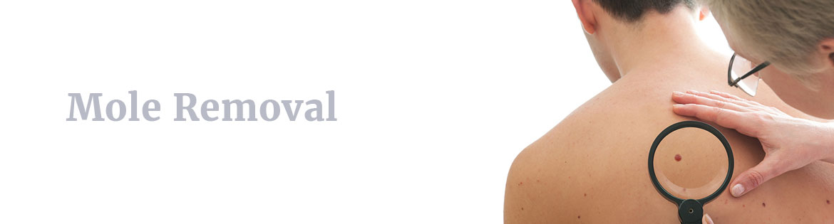
The diagnosis of a mole involves a thorough evaluation by a dermatologist, who will assess its appearance, size, and any changes over time. The key to diagnosing whether a mole should be removed lies in the clinical examination, the use of advanced diagnostic techniques, and the patient's medical history.
Steps in Mole Diagnosis
-
Clinical History: The dermatologist will inquire about when the mole appeared, any changes in its appearance, family history of skin cancer, and your general health. This helps in assessing the risk of malignancy.
-
Physical Examination: The mole will be examined closely using a dermatoscope, which magnifies the mole and helps the doctor see its internal structure. The characteristics of the mole, such as its shape, color, and borders, will be carefully noted.
-
Biopsy: If there is any suspicion of skin cancer or atypical features, a biopsy is performed. The most common types of biopsies for mole evaluation include:
-
Excisional biopsy: The mole is completely removed along with a small margin of surrounding tissue for analysis.
-
Shave biopsy: The mole is shaved off at the skin level and sent for testing.
-
Punch biopsy: A small, circular piece of tissue is removed from the mole for further examination.
-
-
Pathological Evaluation: After biopsy, the sample is sent to a laboratory where a pathologist examines the tissue for cancerous cells, such as melanoma, or benign conditions like seborrheic keratosis or dermatofibromas.
Decision to Remove
-
If the mole is confirmed to be benign, the decision to remove it is often based on cosmetic or personal preference.
-
If the mole is atypical or suspicious, removal is necessary to prevent further complications or to facilitate biopsy for a definitive diagnosis.
Treatment Options for Mole Removal - Methods and Approaches
There are several methods for mole removal, and the choice of technique depends on factors such as the mole's location, size, type, and whether malignancy is suspected.
Surgical Excision
Surgical excision is the most common and preferred method for mole removal, especially when cancer is suspected. In this procedure, the mole and a margin of surrounding skin are removed using a scalpel, and the wound is then closed with stitches. This method allows for complete removal of the mole and ensures that the tissue can be sent for pathological examination.
Shave Excision
In shave excision, the mole is shaved off at or just beneath the surface of the skin using a scalpel or a razor-like instrument. This method is typically used for benign moles that do not need deep tissue removal, and it's preferred for smaller, raised moles. However, it may not be suitable for moles that are suspected of being malignant, as it does not provide the full depth of tissue for biopsy.
Laser Removal
Laser removal is a non-invasive technique often used for cosmetic reasons. It involves using a focused beam of light to vaporize the mole tissue. This method is suitable for smaller, flat moles that are not suspected to be cancerous. However, laser removal is not recommended when malignancy is suspected, as it may not provide enough tissue for biopsy.
Cryotherapy
Cryotherapy involves freezing the mole with liquid nitrogen, causing the tissue to die and fall off over time. This method is typically used for benign moles or warts but is less effective for larger or deeper moles. It also does not allow for tissue sampling for pathological examination.
Electrosurgery
Electrosurgery uses a high-frequency electrical current to burn off the mole. Like laser removal, this technique is often used for cosmetic reasons and is suitable for small, raised moles. It can be combined with other treatments, such as excision, if needed.
Each of these methods has its pros and cons, and the appropriate technique will be selected based on the mole's characteristics and the patient's needs. The choice of method will be discussed with the dermatologist, who will ensure that the procedure is performed safely and effectively.
Pre-Surgical, Intra-Surgical, and Post-Treatment Management
Pre-Surgical Management
Before undergoing mole removal, a thorough evaluation is essential to ensure that the procedure is safe and suitable for the patient. This may include:
-
Medical History Review: To assess for any underlying conditions that could affect healing, such as diabetes or bleeding disorders.
-
Assessment of the Mole: The dermatologist will examine the mole, determine if it requires further evaluation, and discuss the type of removal procedure best suited for the situation.
Intra-Surgical Management
During the mole removal procedure:
-
Local Anesthesia is typically administered to numb the area, ensuring the procedure is pain-free.
-
The mole is carefully removed using the chosen method, and the tissue is sent for pathology if necessary.
-
The wound is then cleaned, and if stitches are required, they will be applied.
Post-Treatment Management
-
Wound Care: After the procedure, the wound will need to be kept clean and covered for a few days. Follow the dermatologist's instructions for cleaning the site and changing the dressing.
-
Pain Management: Any mild discomfort can usually be managed with over-the-counter pain relievers.
-
Avoid Sun Exposure: It's important to protect the site from sun exposure to reduce the risk of pigmentation changes or scarring.
-
Follow-Up: A follow-up appointment may be scheduled to remove any stitches (if applicable) and review the pathology results.
Complications of Mole Removal
Though mole removal is generally a safe procedure, there are potential risks and complications associated with it. The most common complications include:
-
Infection: Although rare, infection at the site of removal can occur, especially if the wound is not kept clean.
-
Scarring: While most mole removal methods result in minimal scarring, some patients may develop noticeable scars. The risk of scarring is higher if the mole is large or removed from a visible area like the face.
-
Recurrence: In some cases, a mole may grow back if not completely removed.
-
Hyperpigmentation or Hypopigmentation: After healing, the area where the mole was removed may be darker (hyperpigmentation) or lighter (hypopigmentation) than the surrounding skin, especially in patients with darker skin tones.
-
Nerve Damage: In rare cases, if the mole is close to nerves, removal could lead to temporary numbness or tingling at the site.
Living with the Condition After Mole Removal
Post-procedure care is essential for ensuring proper healing and minimizing complications. Most people recover without issues, but it's important to monitor the site for any signs of infection or abnormal healing.
Recovery Timeline
-
First Few Days: The treated area may be slightly swollen or tender. You may experience some redness around the wound, but this usually subsides after a few days.
-
1-2 Weeks: If stitches are used, they will be removed. The wound should be healing, but it's important to follow any aftercare instructions to avoid infection or irritation.
-
3-6 Months: Scarring may take several months to fully heal. It's important to continue protecting the area from sun exposure to prevent darkening of the scar.
-
Follow-Up Appointments: Regular follow-ups are important to assess the healing process, remove stitches, and review pathology results.
Cosmetic Considerations
For those who have had moles removed for cosmetic reasons, it's essential to understand that some scarring may remain, especially if the mole was large or located on sensitive areas. Scar minimization techniques, such as silicone sheets or gels, can help reduce the appearance of scars over time.
Top 10 Frequently Asked Questions about Removal of Moles
1. What exactly does “mole removal” involve?
Mole removal is a minor surgical procedure performed by a dermatologist or suitably trained surgeon to remove a skin growth (a mole) for either cosmetic reasons, comfort (if it rubs or becomes irritated) or medical reasons (if it looks suspicious). The procedure typically begins with cleaning and numbing the area, then using one of several removal techniques—such as shave excision (shaving off the mole flush with the skin), punch excision (removing a plug of tissue), or full surgical excision (cutting out the mole plus a margin of healthy skin and stitching the wound). After removal, the tissue may be sent to a pathology lab for examination if there is concern about skin-cancer risk.
2 Why would someone choose to have a mole removed?
There are several reasons:
-
Medical concern: If the mole has changed in size, shape, colour, bleeds or causes itching, it could be a warning sign and needs evaluation/removal.
-
Functional discomfort: If a mole is in a spot that rubs against clothing or jewelry, is constantly irritated or hurt by shaving, removal can improve comfort.
-
Cosmetic reasons: Many people remove moles simply because they don't like the way it looks, especially on visible areas like the face, neck or hands.
-
Prevention or vigilance: Although most moles are benign, removal may be considered if the dermatologist suspects an atypical mole.
3. What methods are used for removing moles and how are they chosen?
The choice of removal method depends on the mole's size, depth, location, suspected nature (benign vs suspicious), and the desired cosmetic outcome. Common methods include:
-
Shave excision: Mole is shaved off with a blade just under the skin surface; useful for raised moles that likely are benign.
-
Punch excision: A round blade (punch) removes a “plug” of skin including the mole; useful for moderately sized lesions.
-
Surgical excision (elliptical removal with stitches): The mole plus a margin of healthy skin is removed, then the wound is sutured; used when the mole is deep or there's suspicion of cancer so pathology is needed.
Each method has trade-offs in terms of scarring, healing time and certainty of complete removal.
4. Is the procedure painful?
Because the area is numbed with local anaesthesia, most patients feel very little pain during the removal. They might feel a pinch or pressure as the anaesthetic is injected and then almost nothing during the removal itself. Some mild discomfort, soreness or stinging may occur for a few days afterwards, especially if the area is on a mobile part of the body or subject to movement/irritation.
5. What are the risks or complications of mole removal?
Although mole removal is a routine procedure, there are some risks, including:
-
Bleeding or oozing from the wound site, particularly in areas with many blood vessels.
-
Infection if the wound is not kept clean or if proper aseptic technique is not followed.
-
Scarring, which may be more noticeable depending on the size of the mole, the method used, skin type (for example darker skin tones may have pigment changes) and the body location.
-
Recurrence of the mole: If the mole was not removed completely or if the method used left some cells behind, the mole might regrow.
-
Delayed diagnosis: If a mole that is potentially cancerous is removed only superficially (for example by laser or home removal) and not sent for pathology, the diagnosis of skin cancer may be delayed.
6. How long is the healing period and what should I expect after the procedure?
Healing time depends on the size, location and method of removal. In many cases, the wound will heal in about 1-3 weeks, though full maturation of the scar may take months. For minor removals, you may be back to normal activities quickly. The area will likely form a scab, then the scab falls off, followed by pink/skin-coloured healing tissue which gradually fades. Proper aftercare is key. You may experience mild redness, swelling or discomfort during the early period. You will likely be advised to keep the area clean and moist (ointment) and protected from the sun to reduce pigmentation and scarring.
7 Will there be a scar and how much will it matter?
Yes, in most cases a scar will result, though its prominence varies. If the removal is superficial with minimal invasion, the scar may be very small and fade over time. More extensive removals (deeper excision with stitches) may leave a thin linear scar. The final scar appearance depends on: how well the wound is cared for, how well the skin naturally heals, your skin type (darker skin may have more pigment change), the body region, and sun exposure during healing. Good after-care—keeping the area clean, applying stabilising ointment, sun protection—makes a significant difference.
8. Does mole removal also mean I'm cured of any risk of skin cancer?
Not necessarily. Mole removal when done because of suspicion of skin cancer is followed by pathology (lab analysis of the removed tissue) to determine whether cancer cells are present and whether margins are clear. Even when a mole is benign, new moles may appear or other skin cancers may arise elsewhere. Regular skin checks are still important. If the mole was atypical or high-risk, your dermatologist may recommend closer surveillance or further treatment.
9. Can I remove a mole at home or with over-the-counter products?
No. It is strongly advised not to attempt mole removal at home or by non-medical methods such as home creams, “DIY” cutters, or unapproved laser pens. These methods may leave mole cells behind, misdiagnose a cancerous lesion, cause infection, excessive scarring or other complications. Professional removal ensures sterile technique, appropriate method, and pathology if needed.
10. What after-care should I follow to ensure best healing and results?
After the procedure, you should follow your doctor's or dermatologist's instructions carefully. Key points include:
-
Keep the area clean and gently wash as advised.
-
Apply any prescribed ointment or recommended emollient to keep wound moist, which helps healing.
-
Avoid picking at the scab or disturbing the wound site.
-
Protect the site from sun exposure (use sunscreen once healed enough) to avoid pigmentation changes.
-
Monitor the area for signs of infection (redness, heat, increasing pain, discharge) or for any regrowth of the mole and report to your dermatologist if you see any worrying signs.
-
Attend any follow-up appointment (for suture removal or pathology discussion) as recommended.
