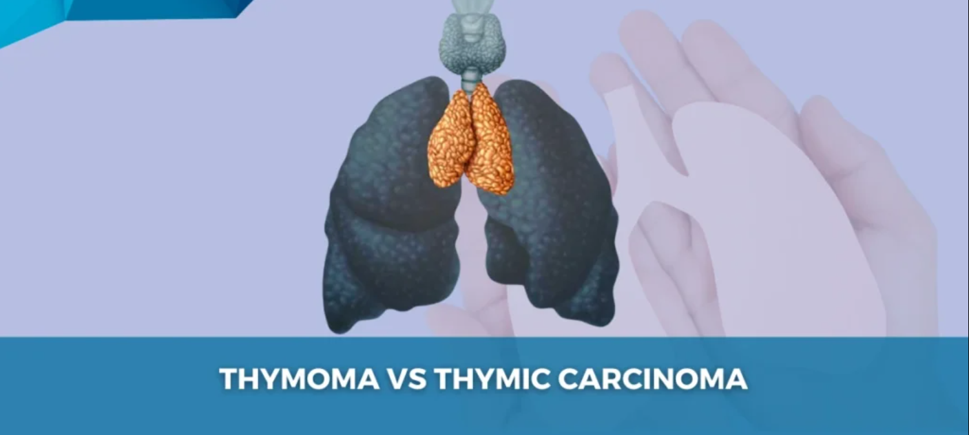
Introduction to Thymoma and Thymic Carcinoma
Thymoma and thymic carcinoma are both rare and complex cancers originating in the thymus gland, a key organ involved in the immune system. Located behind the breastbone in the chest, the thymus plays a critical role in the development of T-lymphocytes (T-cells), which are essential for immune responses.
While thymoma is generally a slow-growing tumor with a better prognosis, thymic carcinoma is a rare, more aggressive cancer that can spread to other organs.
Thymoma Overview
Thymomas are typically benign tumors that arise from the epithelial cells of the thymus. These tumors tend to remain confined to the thymus but can still cause significant symptoms as they grow larger, compressing nearby structures like the lungs, heart, and large blood vessels.
Thymic Carcinoma Overview
Thymic carcinoma is a far more aggressive form of cancer, characterized by rapid growth and the potential for metastasis. It has a higher malignant potential compared to thymomas and often spreads to other areas such as the lungs, lymph nodes, and liver. Early diagnosis and intervention are critical for improving prognosis.
Causes and Risk Factors of Thymoma and Thymic Carcinoma
Although the precise causes of thymoma and thymic carcinoma remain unclear, several genetic and environmental factors are believed to contribute to their development.
Genetic Causes
-
Chromosomal Abnormalities: DiGeorge syndrome, Down syndrome, and multiple endocrine neoplasia (MEN) syndrome are associated with an increased risk of developing thymic tumors.
-
Inherited Syndromes: Some individuals may inherit genetic mutations that increase their likelihood of developing thymoma or thymic carcinoma, including mutations in tumor suppressor genes or those affecting immune system function.
Environmental Factors
-
Radiation Exposure: People who have received radiation therapy to the chest, especially for other cancers, are at higher risk of developing thymic cancers later in life.
-
Chronic Infections: Long-term infections that lead to inflammation in the chest region may increase the risk of developing thymic tumors.
-
Immune System Dysfunction: Autoimmune diseases, particularly myasthenia gravis, are strongly associated with thymomas.
Autoimmune Diseases and Thymic Tumors
-
Myasthenia Gravis (MG): Approximately 30% of people with thymomas develop MG, an autoimmune condition that causes weakness in voluntary muscles. The link between autoimmunity and thymoma is still under investigation.
Symptoms and Signs of Thymoma and Thymic Carcinoma
The symptoms of thymoma and thymic carcinoma depend largely on the tumor's size, location, and its impact on surrounding structures.
Symptoms of Thymoma
-
Chest Pain: Thymomas may cause dull, persistent chest pain, especially when they compress the heart or lungs.
-
Cough: Chronic, non-productive cough, often associated with difficulty breathing.
-
Shortness of Breath: Especially with exertion, or when the tumor compresses the lungs.
-
Hoarseness: Changes in the voice due to pressure on the recurrent laryngeal nerve.
-
Fatigue: A general feeling of weakness and low energy.
-
Difficulty Swallowing: Known as dysphagia, caused by pressure on the esophagus.
-
Swelling in the Face and Neck: Due to superior vena cava syndrome, a condition caused by obstruction of the superior vena cava by the tumor.
Symptoms of Thymic Carcinoma
-
Severe Chest Pain: Thymic carcinoma often causes severe chest discomfort and pressure.
-
Rapid Breathing: Difficulty breathing, particularly if the tumor causes fluid buildup in the lungs.
-
Weight Loss: Unexplained weight loss and loss of appetite.
-
Systemic Symptoms: Symptoms like fever and night sweats may occur if the tumor spreads to other organs.
Paraneoplastic Syndromes
-
Myasthenia Gravis: As mentioned earlier, thymomas are commonly associated with MG, which leads to symptoms like muscle weakness and fatigue.
-
Other Syndromes: Thymomas may also be linked to pure red cell aplasia (a blood disorder) and Good syndrome (a form of immunodeficiency).
Diagnosis of Thymoma and Thymic Carcinoma
Diagnosing thymoma and thymic carcinoma involves various imaging studies and histological examinations.
Imaging Techniques
-
Chest X-ray: The first diagnostic step when a mediastinal mass is suspected. It can show enlarged thymus or signs of compression on adjacent structures.
-
CT Scan: The computed tomography (CT) scan provides highly detailed images that help assess tumor size, shape, and its relationship to surrounding organs.
-
MRI: Magnetic resonance imaging provides high-resolution imaging, especially useful for assessing vascular involvement and the extent of the tumor.
-
PET Scan: A positron emission tomography (PET) scan can be used to detect areas of active tumor and potential metastasis.
Biopsy and Histological Examination
-
Fine-Needle Aspiration (FNA): A thin needle is used to extract a small sample from the tumor for examination.
-
Core Biopsy: A larger tissue sample is taken to provide a more detailed analysis of the tumor's structure.
-
Histopathology: A biopsy is reviewed under a microscope to determine the type of tumor, its grade, and whether it is a thymoma or thymic carcinoma.
Molecular Testing
-
Gene Expression Profiling: In advanced cases, molecular analysis may be used to examine genetic mutations and predict the tumor's behavior and response to treatment.
Treatment Options for Thymoma and Thymic Carcinoma
The treatment of thymoma and thymic carcinoma depends on the tumor's type, stage, and whether it has spread to other areas.
1. Surgical Treatment
-
Thymectomy: The primary treatment for thymomas, thymectomy involves the surgical removal of the thymus gland. For thymic carcinoma, more extensive surgery may be required to remove the tumor and surrounding tissues.
-
Extended Thymectomy: In some cases, nearby lymph nodes and structures are also removed if the tumor has spread locally.
-
2. Radiation Therapy
-
Postoperative Radiation: Radiation therapy is often used after surgery to ensure any remaining cancerous cells are eliminated.
-
Palliative Radiation: In cases where the cancer is inoperable or has metastasized, radiation can be used to alleviate pain, swelling, and other symptoms.
3. Chemotherapy
-
Chemotherapy is often used for thymic carcinoma due to its aggressive nature. Common chemotherapeutic agents include:
-
Cisplatin
-
Doxorubicin
-
Cyclophosphamide
-
Chemotherapy can help manage advanced or metastatic thymic carcinoma and improve overall survival in patients with aggressive disease.
4. Immunotherapy
-
Immune Checkpoint Inhibitors: Drugs such as nivolumab and pembrolizumab are increasingly being tested in clinical trials for the treatment of thymic carcinoma. These drugs work by enhancing the body's immune system to fight the tumor.
5. Targeted Therapy
-
Targeted therapies focus on specific pathways involved in the growth of thymic tumors. Drugs targeting tumor growth factors or blood supply to the tumor are being explored in clinical trials.
Prevention and Management of Thymoma and Thymic Carcinoma
While prevention of thymoma and thymic carcinoma is not entirely possible, there are key strategies for early detection and management:
Prevention
-
Genetic Counseling: Families with a history of congenital conditions like DiGeorge syndrome should consider genetic counseling.
-
Avoid Radiation: Limiting exposure to radiation, particularly in children, can reduce the risk of developing thymic tumors.
Management After Treatment
-
Regular Follow-ups: Patients should be regularly monitored with imaging studies (CT scans, MRIs) to detect recurrence of the disease.
-
Manage Complications: Address any autoimmune conditions or side effects caused by treatment, including radiation therapy and chemotherapy.
Complications of Thymoma and Thymic Carcinoma
Even after treatment, there are several complications associated with thymoma and thymic carcinoma:
-
Recurrence: Thymic carcinoma, in particular, has a higher risk of recurrence due to its aggressive nature.
-
Metastasis: Thymic carcinoma can spread to the lungs, liver, bones, and other distant organs.
-
Superior Vena Cava Syndrome: Compression of the superior vena cava by the tumor can lead to swelling in the upper body and difficulty breathing.
-
Pulmonary Hypertension: Due to compromised lung function, some patients may develop high blood pressure in the lungs.
Living with Thymoma and Thymic Carcinoma
While thymoma and thymic carcinoma are challenging conditions, effective treatment allows many patients to lead fulfilling lives after recovery.
Post-Surgery Recovery
-
Physical Rehabilitation: After surgery or treatment, physical rehabilitation can help patients regain strength and improve their quality of life.
Psychosocial Support
-
Mental Health: Coping with a cancer diagnosis can be emotionally draining. Patients and families are encouraged to seek support through counseling or support groups.
Lifestyle Modifications
-
Diet and Exercise: Maintaining a healthy diet and engaging in regular physical activity can improve overall well-being.
Top 10 Frequently Asked Questions about Thymoma and Thymic Carcinoma
1. What are Thymoma and Thymic Carcinoma?
Thymoma and thymic carcinoma are rare cancers originating in the thymus gland, located behind the breastbone. Thymomas are typically slow-growing and less likely to spread, whereas thymic carcinomas are more aggressive, grow rapidly, and have a higher tendency to metastasize.
2. What are the symptoms of Thymoma and Thymic Carcinoma?
Early-stage thymic tumors often present no symptoms. When symptoms occur, they may include:
-
Persistent cough
-
Chest pain or pressure
-
Shortness of breath
-
Difficulty swallowing
-
Hoarseness
-
Swelling of the face, neck, or arms
-
Muscle weakness
-
Anemia
-
Frequent infections
-
Unexplained weight loss
These symptoms can also be indicative of other conditions, so it's essential to consult a healthcare professional for an accurate diagnosis.
3. How are Thymoma and Thymic Carcinoma diagnosed?
Diagnosis typically involves:
-
Imaging tests: Chest X-ray, CT scan, MRI, or PET scan to identify abnormalities.
-
Biopsy: A tissue sample is examined to confirm the presence of cancer cells.
-
Blood tests: To detect associated autoimmune conditions or other abnormalities.
Early detection often occurs incidentally during imaging for other issues.
4. What is the difference between Thymoma and Thymic Carcinoma?
Thymoma cells resemble normal thymus cells, grow slowly, and rarely spread. In contrast, thymic carcinoma cells appear abnormal, grow rapidly, and are more likely to metastasize .
5. What are the treatment options for Thymoma and Thymic Carcinoma?
Treatment strategies depend on the tumor's type, stage, and resectability:
-
Surgery: Primary treatment to remove the tumor.
-
Radiation Therapy: Used post-surgery or for inoperable tumors.
-
Chemotherapy: Administered before surgery (neoadjuvant) or after
(adjuvant) to shrink or eliminate cancer cells.
-
Targeted Therapy: Drugs that target specific cancer cell
mechanisms.
-
Hormone Therapy: For certain thymomas responsive to hormones.
-
Immunotherapy: Under investigation for advanced cases.
6. What is the prognosis for individuals with Thymoma and Thymic
Carcinoma?
The prognosis varies:
-
Thymoma: Generally favorable with a 5-year survival rate of
approximately 90% .
-
Thymic Carcinoma: More aggressive with a 5-year survival rate
ranging from 36% to 55%, depending on the stage and treatment response.
7. Who is at risk for developing Thymoma and Thymic Carcinoma?
Thymomas are more common in adults aged 40-75, particularly among Asian and Pacific
Islander populations. Thymic carcinoma is rarer and often diagnosed at advanced stages.
Risk factors include genetic conditions like MEN1 (Multiple Endocrine Neoplasia type 1)
and associations with autoimmune diseases.
8. Can Thymoma be associated with other health conditions?
Yes, thymoma is commonly linked with autoimmune paraneoplastic syndromes, where the
immune system attacks normal tissues. The most common is myasthenia gravis, a
neuromuscular disorder.
9. Is there a staging system for Thymoma and Thymic Carcinoma?
Yes, the Masaoka-Koga staging system is widely used, classifying tumors from Stage I
(localized) to Stage IV (metastatic). This system helps determine the extent of disease
and guides treatment planning.
10. What is the role of follow-up care after treatment?
Regular follow-up is crucial to monitor for recurrence or complications. This may
include imaging tests, physical exams, and assessments for associated autoimmune
conditions. Lifelong monitoring is often recommended, especially for thymoma patients.
Surgery: Primary treatment to remove the tumor.
Radiation Therapy: Used post-surgery or for inoperable tumors.
Chemotherapy: Administered before surgery (neoadjuvant) or after (adjuvant) to shrink or eliminate cancer cells.
Targeted Therapy: Drugs that target specific cancer cell mechanisms.
Hormone Therapy: For certain thymomas responsive to hormones.
Immunotherapy: Under investigation for advanced cases.
Thymoma: Generally favorable with a 5-year survival rate of approximately 90% .
Thymic Carcinoma: More aggressive with a 5-year survival rate ranging from 36% to 55%, depending on the stage and treatment response.


